
|
Fig. S2
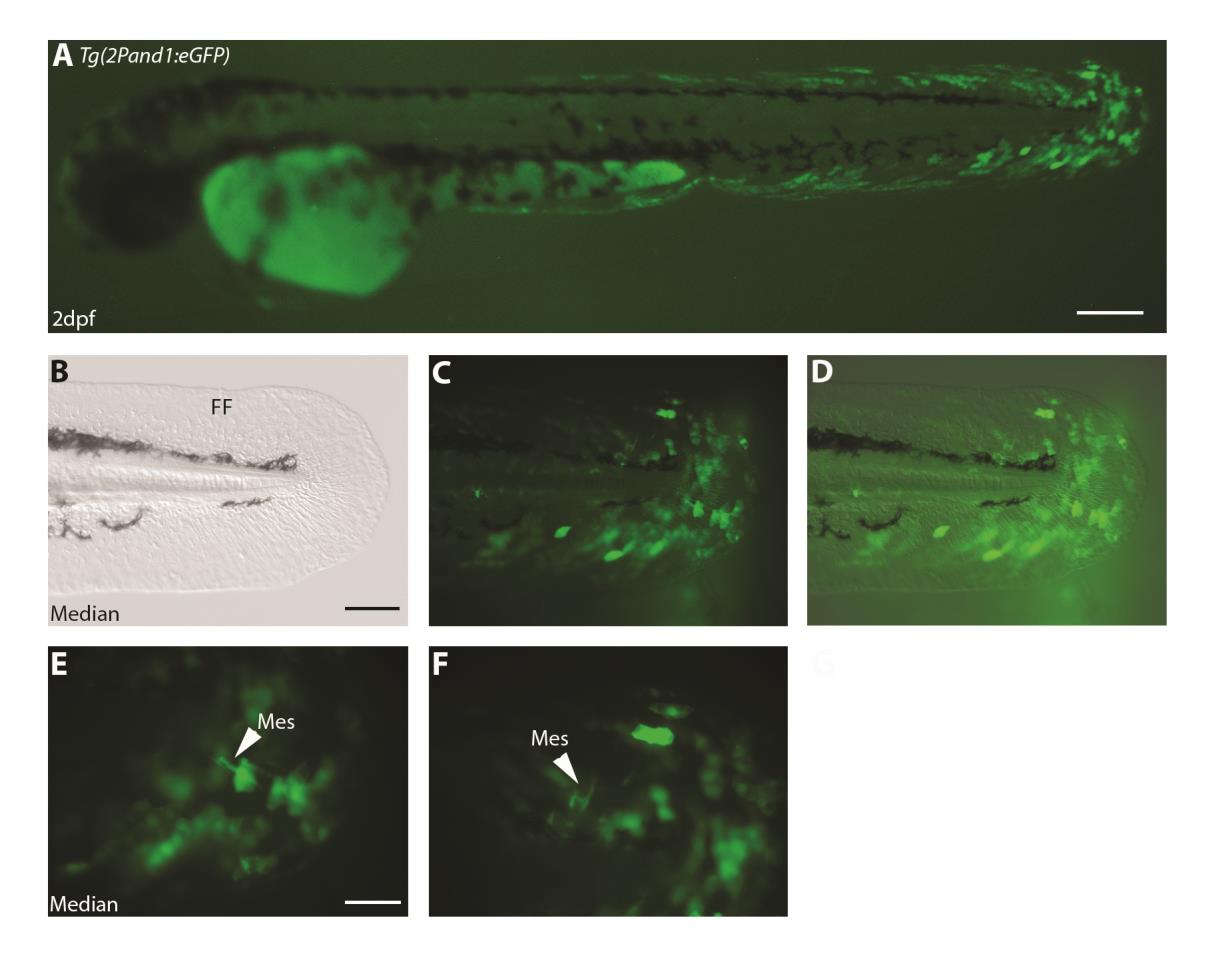
Mosaic ectodermal expression in Tg(2Pand1:eGFP) injected embryos reveal mesenchymal expression. (A-F) Tg(2Pand1:eGFP) transient expression in injected embryos at 2 d.p.f.. Following injection of a DNA construct in zebrafish embryo, expression of the transgene is always mosaic. This allows to visualize GFP expression in some mesenchymal cells (indicated by white arrowheads) due to the mosaic GFP expression in the overlaying ectodermal cells (E, F). Brightfield (B), fluorescence (A, C, E, F), and brightfield/fluorescence merged images (D). FF, Fin fold; Epi , Ectodermal cells; Mes, Mesenchymal cells. Scale bars: 200µm in A; 100µm in B-D; 50µm in E, F.
Reprinted from Developmental Biology, 417(1), Lalonde, R.L., Moses, D., Zhang, J., Cornell, N., Ekker, M., Akimenko, M.A., Differential actinodin1 regulation in zebrafish and mouse appendages, 91-103, Copyright (2016) with permission from Elsevier. Full text @ Dev. Biol.