
|
Fig. 8
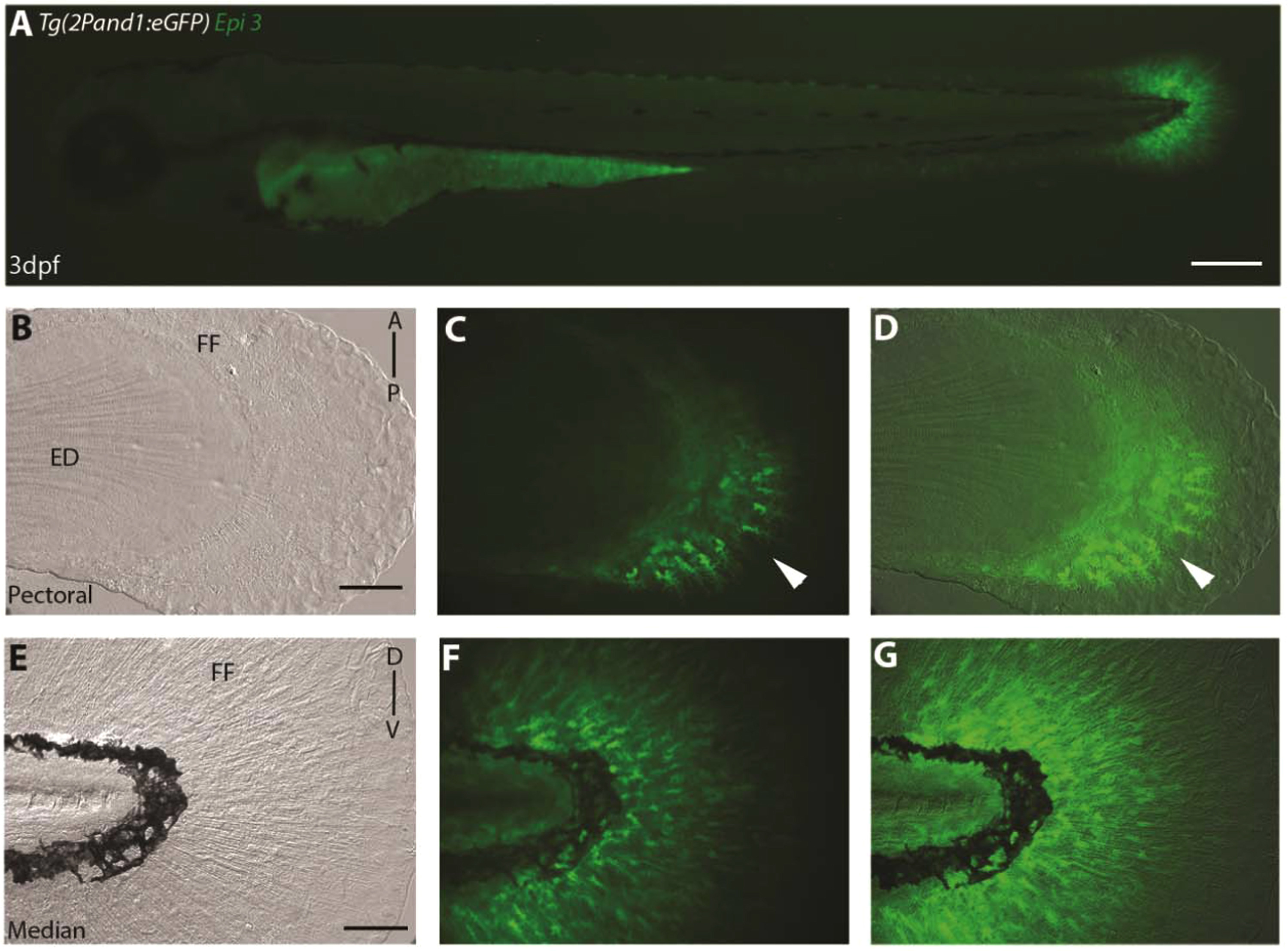
Epi site 3 required for epithelial reporter expression in Tg(2Pand1:eGFP). (A-G) 3 d.p.f. Tg(2Pand1:eGFP) epi site 3 transgenic reporter line. Ectodermal expression is completely lost with the removal of the epi site 3 23 b.p. fragment (A-G). Median (E-G) and pectoral fin fold (B-D) display reporter expression in the mesenchymal cells identical to Tg(2PΔepi:eGFP) transgenic line. Note the higher number of eGFP-positive cells in the posterior half of the pectoral fin fold (B, C). Brightfield (B, E), fluorescence (A, C, F), and brightfield/fluorescence merged images (D, G). ED, Endoskeletal Disc; FF, Fin Fold; A, Anterior; P, Posterior; D, Dorsal; V, Ventral. Scale bars: 200 µm in A; 50 µm in B-G.
Reprinted from Developmental Biology, 417(1), Lalonde, R.L., Moses, D., Zhang, J., Cornell, N., Ekker, M., Akimenko, M.A., Differential actinodin1 regulation in zebrafish and mouse appendages, 91-103, Copyright (2016) with permission from Elsevier. Full text @ Dev. Biol.