
|
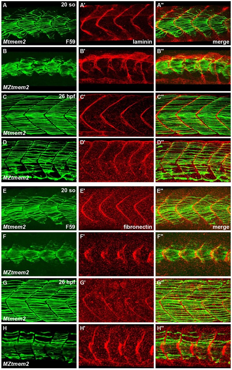
Fig. 2
Aberrant ECM organization at the MTJ in MZtmem2 mutants. (A-H) Immunofluorescence indicates localization of laminin (red, A′-D′) and fibronectin (red, E′-H′) relative to slow muscle fibers, labeled with F59 (green, A-H); lateral views, dorsal up, at 20 somite stage (so) (A,B,E,F) and 26hpf (C,D,G,H). (A-D) Laminin is present at the MZtmem2 MTJ by 20 so (B), although it appears disorganized compared with localization in Mtmem2 siblings (A). By 26hpf, laminin deposition appears diminished at the MZtmem2 MTJ (D). (E-H) Fibronectin fibrillogenesis is evident at the MZtmem2 MTJ at 20 so (F), albeit in an aberrant pattern that echoes the morphology of the MZtmem2 somites. By 26hpf, when much of the fibronectin has been degraded at the Mtmem2 MTJ (G), fibronectin levels appear increased in MZtmem2 mutants (H), particularly where fibers are detached. However, nearly all of this fibronectin degrades in MZtmem2 mutants by 40hpf (data not shown).