
|
Fig. 1
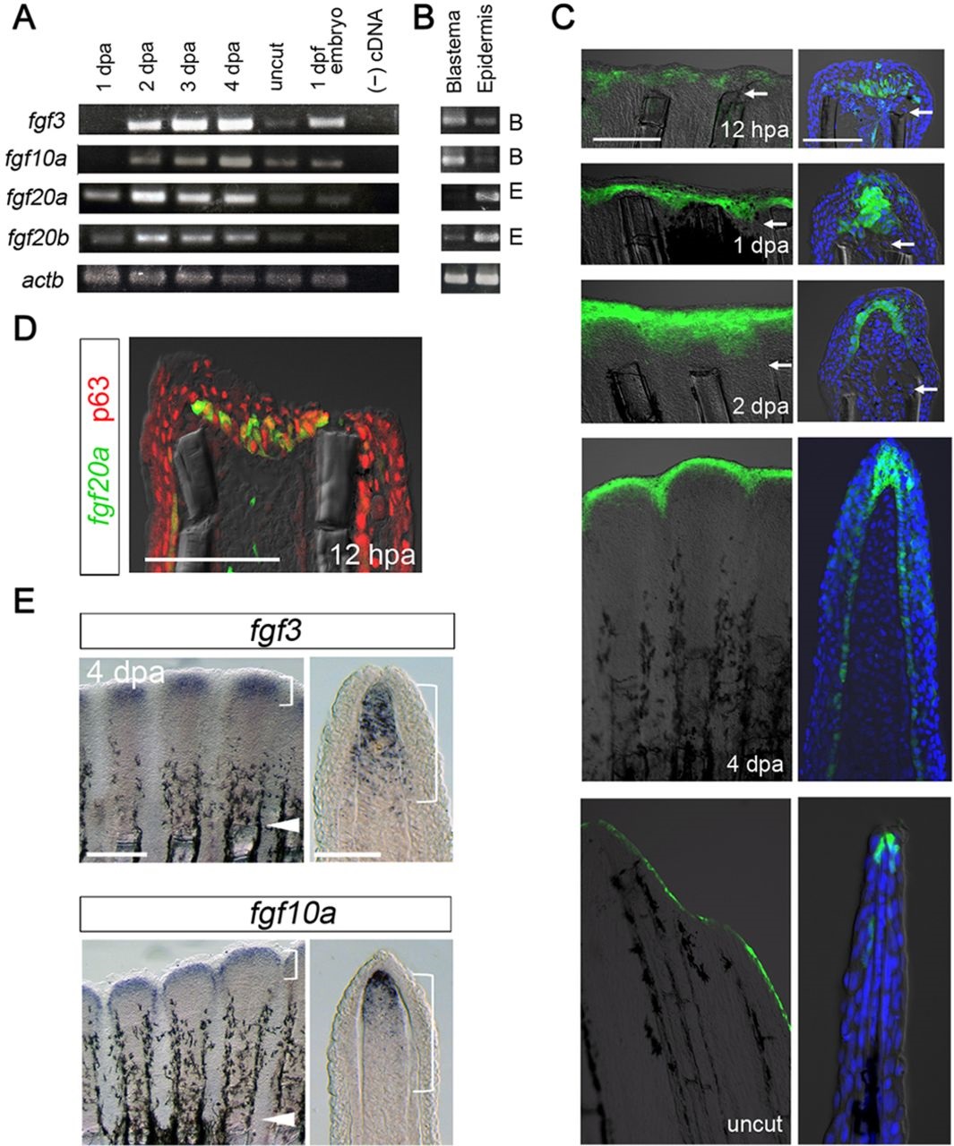
The expression of Fgf ligands during zebrafish fin regeneration. (A) RT-PCR analysis of the expression of fgf3, fgf10a, fgf20a and fgf20b at 1, 2, 3 or 4dpa and in uncut fins. The presented data is a part of comprehensive analysis shown in Fig. S1. The number of PCR cycles was 30. actb1, β-actin1 primers. (B) RT-PCR analysis of tissue-specific expression of Fgf genes in the blastema and wound epidermis at 2dpa. The number of PCR cycles was 35. Fgf genes that are mainly expressed in the blastema or wound epidermis are indicated as B (blastema) or E (epidermis) to the right of the panel. (C) Localisation of fgf20a expression in the wound epidermis, as visualised by the enhancer trap line, HGn21A. Enhanced green fluorescent protein (EGFP)-positive cells gave rise to the basal epidermal cells at 2dpa. In the ‘no-regeneration’ state (uncut fin), EGFP was detected in the distal tip of the epidermis. Tissue sections (right panels) were counter-stained with 4′,6-diamidino-2-phenylindole (DAPI). Arrows indicate the site of amputation. Scale bars: 200µm for whole-mount analysis (left panels); 50µm for sections (right panels). (D) Initiation of fgf20a expression in the epidermal cells. A cryostat section of HGn21A regenerate at 12hpa stained with the antibody against p63, a marker of epidermal cells. Scale bar: 50µm. (E) Whole-mount in situ hybridisation (ISH) analysis of fgf3 and fgf10a expression at 4dpa (left panels) and their respective tissue sections (right panels). Expression of these Fgf genes was strongest in the distal region of the blastema, though the region of fgf3 expression was broader than that of fgf10a. Arrowheads mark amputation sites; brackets indicate approximate areas of the blastema. Scale bars: 200µm (left); 50µm (right).