
|
Fig. S12
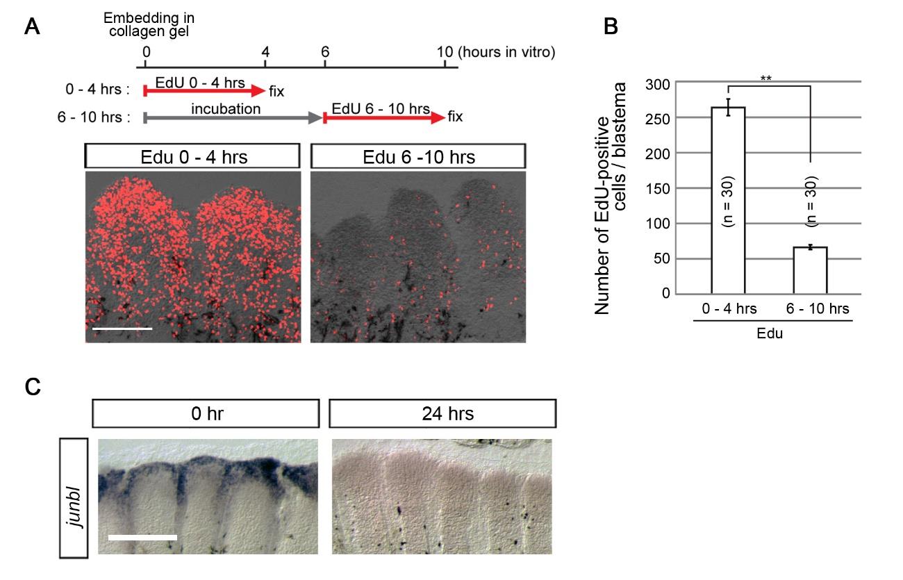
Cell proliferation was not maintained in blastema explants without the wound epidermis. (A) EdU incorporation in blastema explants that were detached from the wound epidermis and incubated in vitro in the L15 medium for 4 hrs (left panel) or 10 hrs (right panel). The Edu labelling was performed for 4 hrs before fixation. Cell proliferation of blastemal explants dramatically decreased within a few hours. Scale bar, 200µm. (B) Quantification of EdU incorporation in the blastema explant. Error bars represent mean ± s.e.m. Student’s t test was performed to assess statistical significance. **p < 0.001. n, the number of blastema, which were taken from 4 fish. (C) Whole-mount ISH analysis of junbl, a distal blastema marker, in blastema explants that were incubated in vitro for 0 hr (n = 5) or 24 hrs (n= 5). The junbl expression rapidly diminished with time and disappeared by 24 hrs. Scale bar, 200 µm.