
|
Fig. 4
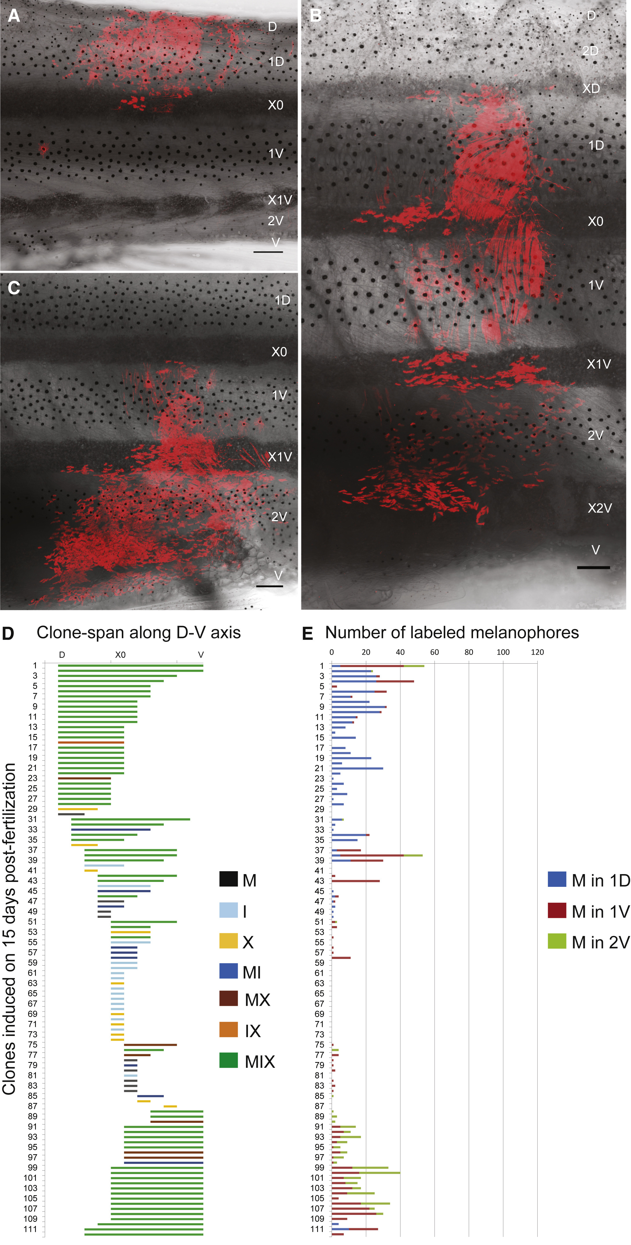
Regional Distribution of Clonally Derived Pigment Cells
(A-D) Clones in young adult animals obtained from Cre activation at 15 dpf (genotype Tg(sox10:ERT2-Cre)/+; Tg(βactin2:loxP-STOP-loxP-DsRed-express)/+) show variability in clone span along the dorsoventral body axis: the clonally derived pigment cells may predominantly contribute to pigment cells of the (A) dorsal, (B) lateral, or (C) ventral regions. Scale bars represent 250 µm. (D) Quantification of the extent of the clones along the dorsoventral body axis; each line represents the dorsoventral span of a single clone. Color code indicates clone type according to pigment cell composition. MIX clones tend to be larger than clones containing only a single pigment cell type. D, dorsal; V, ventral; X0, first light stripe.
(E) Quantification of the number of labeled melanophores in each clone; each line represents the number of melanophores in an individual clone. Color code indicates the dark stripe: blue, 1D; brown, 1V; green, 2V.
Reprinted from Developmental Cell, 38(3), Singh, A.P., Dinwiddie, A., Mahalwar, P., Schach, U., Linker, C., Irion, U., Nüsslein-Volhard, C., Pigment Cell Progenitors in Zebrafish Remain Multipotent through Metamorphosis, 316-30, Copyright (2016) with permission from Elsevier. Full text @ Dev. Cell