
Image
|
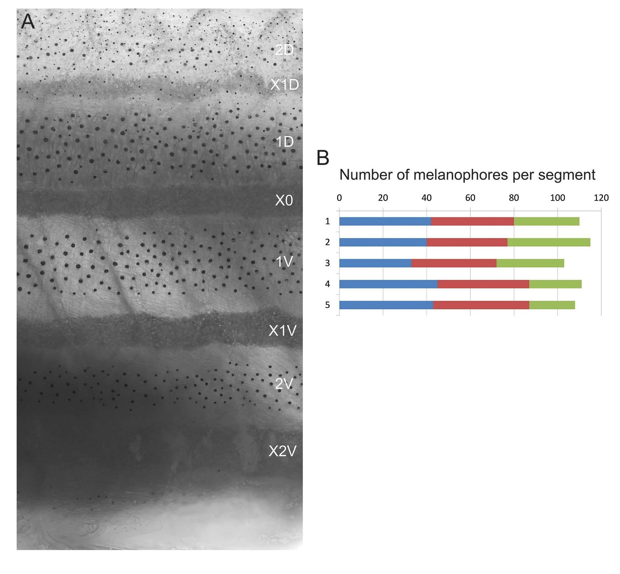
Figure Caption
Fig. S4
Melanophore quantification in wild-type.
(A) Skin of a wild-type young adult zebrafish (22 mmSL, 81 dpf). (B) Graph showing number of melanophores per segment from five different animals. Color code indicates the dark stripe: blue - 1D; brown - 1V; green - 2V Melanophores were counted in the region between the pectoral fin and the anal fin.
Acknowledgments
This image is the copyrighted work of the attributed author or publisher, and
ZFIN has permission only to display this image to its users.
Additional permissions should be obtained from the applicable author or publisher of the image.
Reprinted from Developmental Cell, 38(3), Singh, A.P., Dinwiddie, A., Mahalwar, P., Schach, U., Linker, C., Irion, U., Nüsslein-Volhard, C., Pigment Cell Progenitors in Zebrafish Remain Multipotent through Metamorphosis, 316-30, Copyright (2016) with permission from Elsevier. Full text @ Dev. Cell