
Image
|
Figure Caption
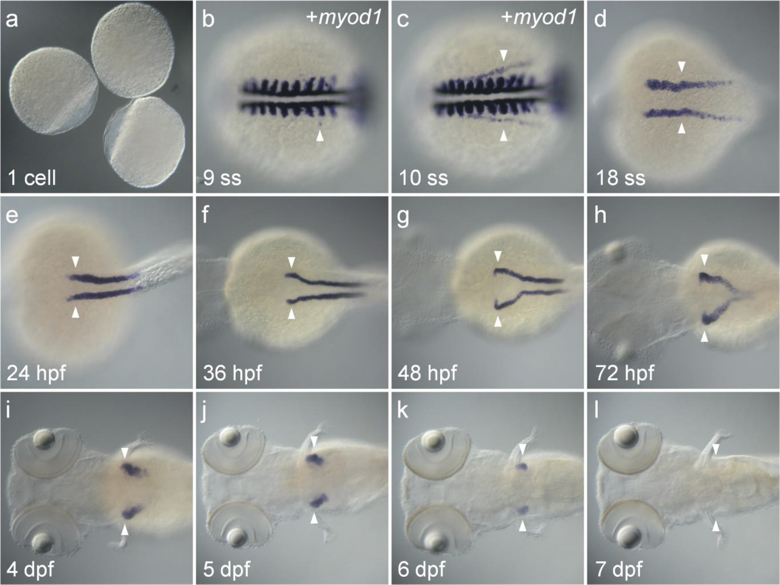
Fig. 5
Pronephros-specific expression pattern of zaqp8b was analyzed by whole mount in situ hybridization. Dorsal view to the left (b-l). Staining without myod1 RNA probe (a, d-l) or co-staining with myod1 probe (b, c). (b) In 9 ss embryos, zaqp8b began to express in a subset of cells in IM (arrowhead). (c-g) Bilateral expression of zaqp8b in pronephros (arrowhead). (h-k) Expression of zaqp8b in the PCT (arrow head) from 72 hpf to 6 dpf. (l) The zaqp8b transcripts in the PCT almost completely disappeared at 7 dpf.
Figure Data
Acknowledgments
This image is the copyrighted work of the attributed author or publisher, and
ZFIN has permission only to display this image to its users.
Additional permissions should be obtained from the applicable author or publisher of the image.
Reprinted from Gene expression patterns : GEP, 21(1), Koun, S., Kim, J.D., Rhee, M., Kim, M.J., Huh, T.L., Spatiotemporal expression pattern of the zebrafish aquaporin 8 family during early developmental stages, 1-6, Copyright (2016) with permission from Elsevier. Full text @ Gene Expr. Patterns