
|
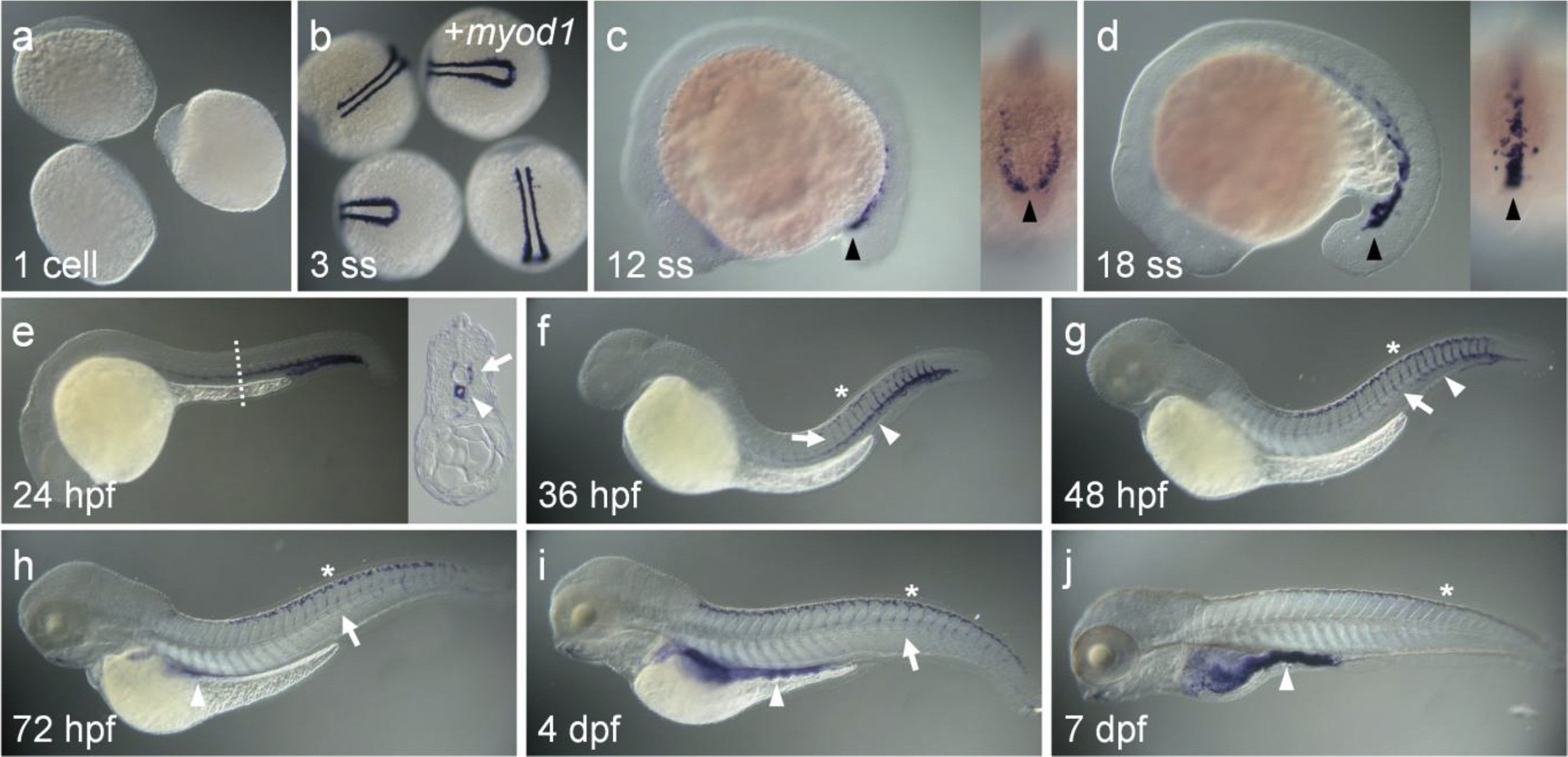
Fig. 2
Expression of zaqp8a.1 in zebrafish embryos was analyzed by whole mount in situ hybridization. Lateral view with anterior to the left (c-j). (a, b) The expression of zaqp8a.1 was not detected at the one cell stage or at 3 ss. For selection of 3 ss embryos precisely, embryos were co-stained with myod1 (b). (c-d) The expression of zaqp8a.1 was first detected in the posterior region of LPM at 12 ss and then anteriorly expanded at 18 ss (arrowhead). (e-j) The expression of zaqp8a.1 was detected in blood vessels. (e) In DA (arrowhead) and ISV (arrow), expression of zaqp8a.1 was seen at 24 hpf, but it greatly decreased in DA and ISV at 72 hpf (h) and 4 dpf (i), respectively. (f) Expression of zaqp8a.1 in DLAV (asterisks) was firstly seen from 36 hpf, and persisted until 7 dpf (j).
Reprinted from Gene expression patterns : GEP, 21(1), Koun, S., Kim, J.D., Rhee, M., Kim, M.J., Huh, T.L., Spatiotemporal expression pattern of the zebrafish aquaporin 8 family during early developmental stages, 1-6, Copyright (2016) with permission from Elsevier. Full text @ Gene Expr. Patterns