
|
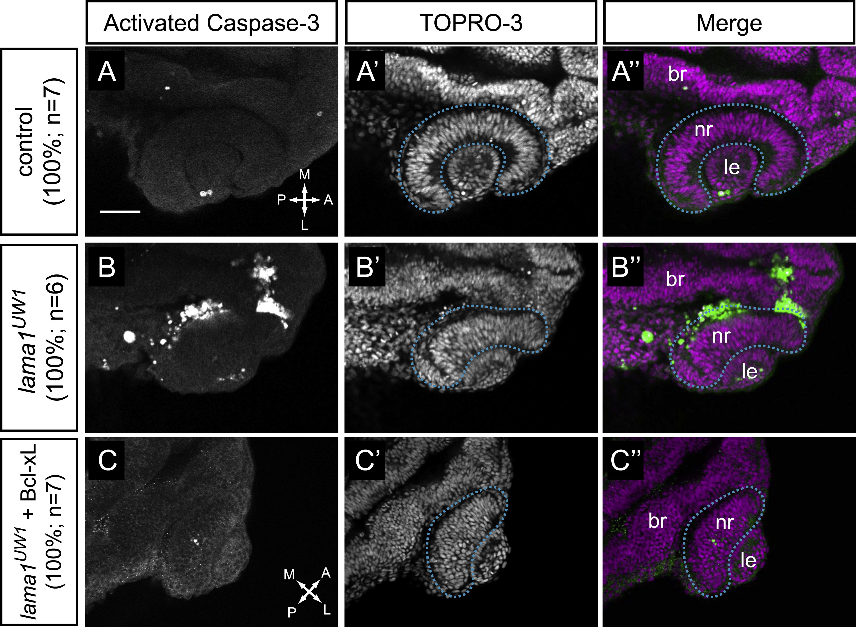
Fig. 4
Apoptosis is increased in lama1UW1 mutant embryos but is not the underlying cause of morphogenesis defects. (A-A′′) Control embryos show little apoptotic cell death. (B-B′′) lama1UW1 mutant embryos contain a significant number of dying cells. (C-C′′) Injection of Bcl-xL RNA (100 pg) rescues apoptosis in lama1UW1 mutant embryos, however optic cup morphogenesis defects are still apparent. (A,B,C) Antibody staining for activated caspase-3. (A′,B′,C′) TOPRO-3 counterstain for nuclei. (A′′, B′′, C′′) Merged images. dashed blue line, boundary of optic cup. Dorsal views; scale bar, 50 µm. br, brain; nr, neural retina; le, lens. A, anterior; P, posterior; M, medial; L, lateral.
Reprinted from Developmental Biology, 416(2), Bryan, C.D., Chien, C.B., Kwan, K.M., Loss of laminin alpha 1 results in multiple structural defects and divergent effects on adhesion during vertebrate optic cup morphogenesis, 324-37, Copyright (2016) with permission from Elsevier. Full text @ Dev. Biol.