
|
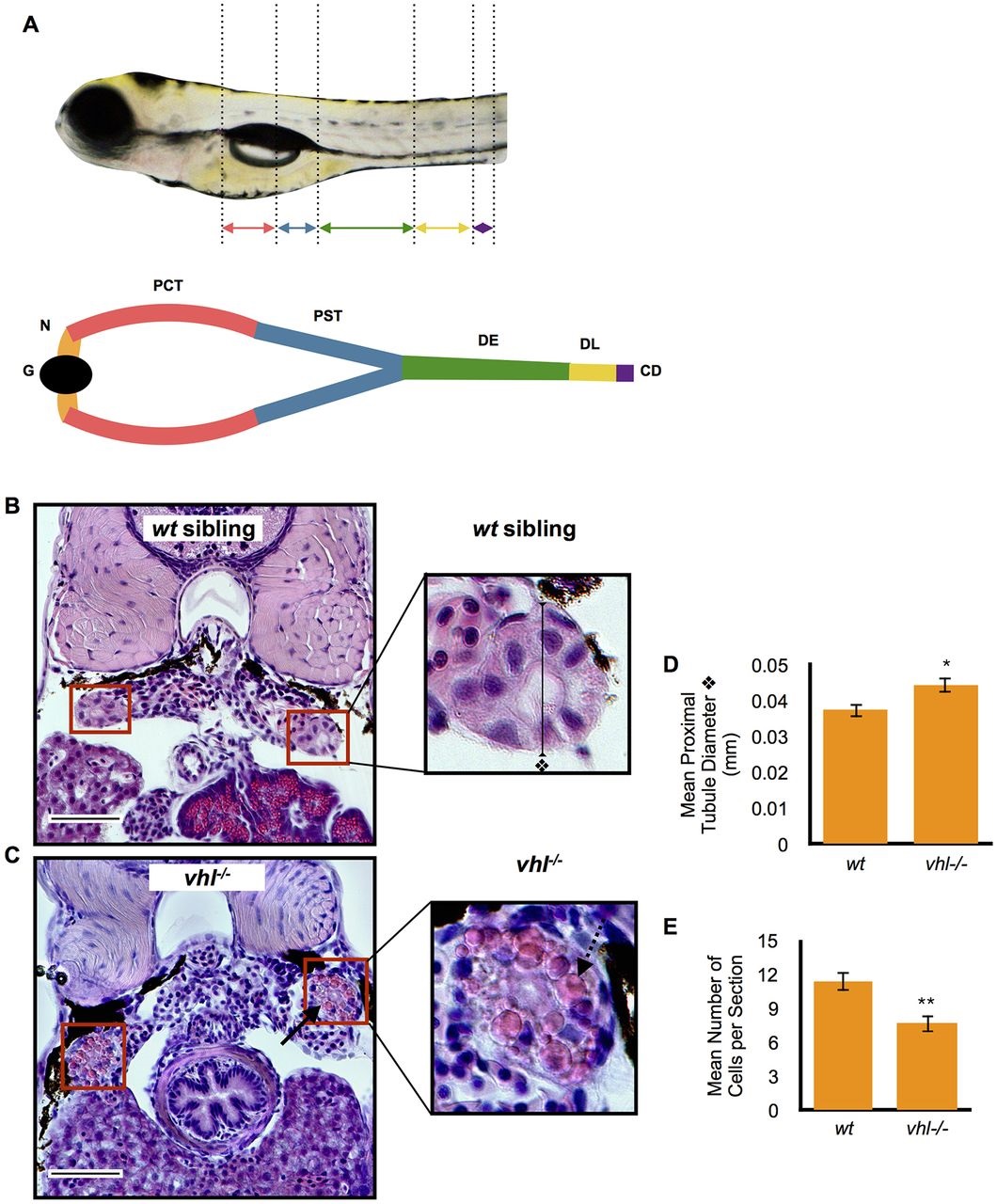
Fig. 1
Proximal vhl-/- pronephric tubules have distorted architecture and contain cytoplasmic vesicles when compared to wt siblings. (A) Location of the proximal and distal pronephric tubules in the zebrafish larval pronephric kidney. Kidney segments are color coded. The glomerulus (G, black), neck (N, orange), proximal convoluted tubule (PCT, red), proximal straight tubule (PST, blue), distal early (DE, green), distal late (DL, yellow), collecting duct (CD, purple) are highlighted. (B,C) 5.5dpf vhl-/- and wt siblings were fixed and embedded in JB-4 and sectioned with a glass knife. H&E staining was performed and measurements were taken using ImageJ. H&E staining of wt sibling (B) and vhl-/- (C) proximal pronephric tubules (red boxes). Scale bars: 0.05mm. (D) Diameter (mm) of proximal pronephric tubule in the transverse section. (E) Mean number of cells per transverse section. Each experiment had at least three larvae per sample group and was performed in duplicate. Data represent mean±s.e.m. *P<0.05, **P<0.01 (paired two-tailed t-test).