
|
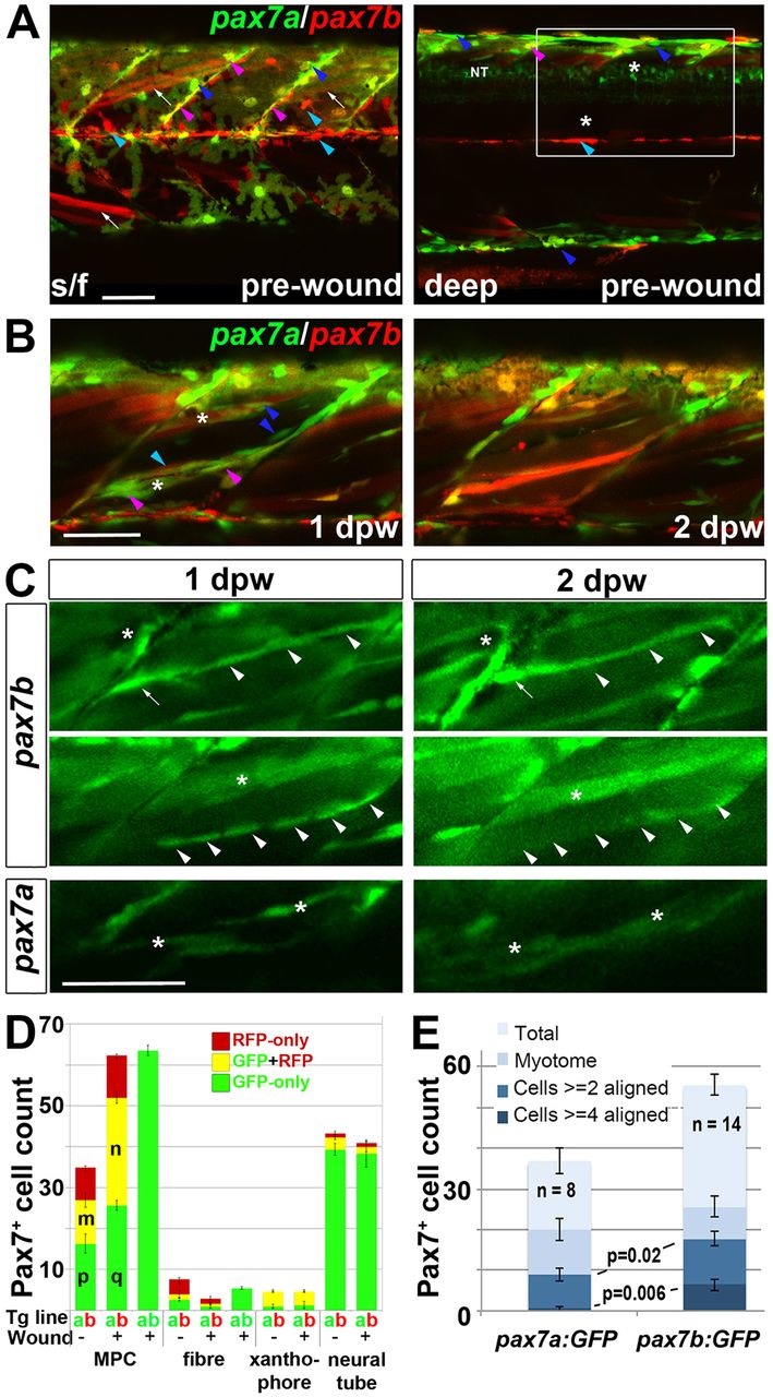
Fig. 5
Fusion of pax7a- and pax7b-reporter cells during wound repair. (A-C) Lateral confocal maximum intensity projection stacks of pre-wounded (A) and wounded (B,C) yolk extension somites of pax7a:GFP;pax7b:gal4;UAS:RFP (A,B) or single pax7a/b:GFP (C) larvae, anterior to left, dorsal to top. Scale bars: 50µm. (A) At 3dpf, pax7b:RFP fibres (white arrows) and presumptive mononucleate cells (cyan arrowheads) are present superficially (s/f) within the somite and differ from pax7a:GFP cells (blue arrowheads). Dual-labelled somite cells (magenta arrowheads) concentrate on VMZ. Note the lack of Pax7 cells in the deep myotome at this stage. The pax7b-reporter labelled cells strongly in somites, and also weakly in dorsal neural tube (NT). (B) Short stack of epaxial wounded region shown by white box in A with two small wounds (asterisks). At 1dpw, pax7a:GFP;pax7b:RFP cells elongate in wound. By 2dpw, time-lapse reveals several nascent fibres marked strongly by RFP and weakly by GFP. See Fig. S9 for separate monochrome images. (C) Time-lapse of pax7b:gal4;UAS:GFP reporter marks aligned cells (arrowheads) that form fibres (top) or disappear (centre). pax7a:GFP cells are frequently aligned with fibres, but more rarely assemble in rows. pax7a:GFP cells occasionally matured into nascent fibres (bottom). Asterisks mark the same cells at each time point. Note the stronger mononucleate cells and more abundant fibre labelling by the pax7b:GFP reporter, compared with the pax7b:RFP reporter in panel B. Arrow indicates a separate cell. (D) Counts of numbers (mean±s.e.m.) of red, green and dual-labelled cells in a single epaxial somite (or corresponding length of neural tube) by cell type in larvae transgenic (Tg) for pax7a:GFP (a, green), pax7b:GFP (b, green) or pax7b:RFP (b, red) as indicated by the Tg line letter code and colour. Larvae with (+) or without (-) a wound made at 3dpf were analysed 1dpw, at 4dpf. Note the increase in labelled MPCs, decrease in fibres and constant number of xanthophores and neurons in wounded somites at 1dpw. Letter groups (m,n,p,q) indicate difference at P<0.05 (t-test, n=3). (E) At 1dpw, despite a similar fraction of total cells in myotome, there were more pax7b:GFP reporter cells in rows of two (≥2) or four (≥4) or more aligned cells, compared with pax7a:GFP cells. Mean±s.e.m., P-values show Mann-Whitney test of differences in proportions of total cells.