
|
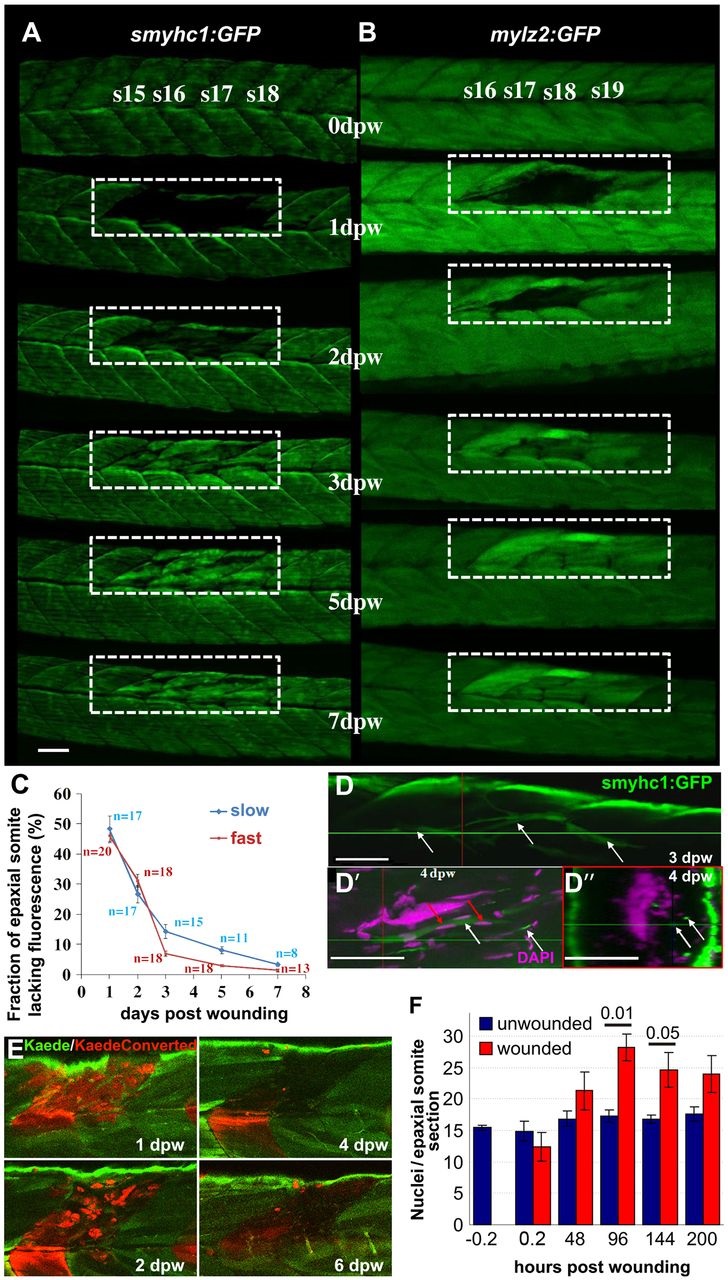
Fig. 1
Time-course of muscle wound repair. (A,B) Large needle incision wounds (boxed regions) in the indicated somites of zebrafish 3.5dpf larvae carrying transgenes expressed in slow (A; smyhc1:gfp) or (B; mylz2:gfp) fast fibres were repeatedly imaged in the same live fish by confocal fluorescence microscopy over 7dpw. Larvae are shown anterior to left, dorsal up. Note the brighter fluorescence of newly synthesised unbleached GFP in regenerated region. s15-s19, somite 15 to somite 19. (C) Rate of recovery (mean±s.e.m.) of GFP fluorescence in epaxial somite of slow smyhc1:gfp and fast mylz2:gfp muscle of n larvae. (D-D′′) smyhc1:gfp larvae showing slow fibres (white arrows) in deep somite, viewed from dorsal (D; 3dpw) and lateral (D′) and corresponding transversal (D′′) views at 4dpw. The red and green crosshairs indicate planes, red arrows indicate elongated fibre-associated nuclei. (E) To investigate the source of new fibres, two adjacent somites in embryos injected with Kaede RNA were photoconverted from green to red at 2.5dpf, then wounded in the epaxial domain and followed for 6dpw. Representative confocal slices in lateral view show loss of KaedeRed without replacement by KaedeGreen. (F) Loss and gain of nuclei (mean±s.e.m.) in epaxial somites of Tg(h2afva:H2AFVA-GFP)kca66 larvae wounded at 3.5dpf and imaged until 12dpf (ANOVA, n=4). Scale bars: 50µm.