
|
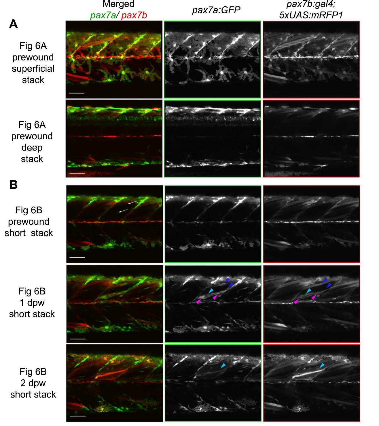
Fig. S9
Individual green and red channels showing fusion of pax7a- and pax7b-reporter cells during wound repair (from Fig. 6A,B). Lateral confocal maximum intensity projection stacks of pre-wound (upper three rows. from Fig. 5A) and post-wound (lower two rows. from Fig. 58) yolk extension somites of a pax7a:GFP;pax7b:RFP larva, anterior to left, dorsal to top. A. At 3 dpf, prior to wounding, xanthophores have strong GFP and weak RFP, neural tube cells have strong GFP and little RFP, whereas superficial muscle fibres have RFP but lack GFP. B. Short stack of epaxial wounded somite region excluding deep and superficial regions. Prior to wounding, no pax7a:GFP cells and only a pax7b:RFP large fibre are present in the deep somite. Two oblique needle insertions made two small lesions in a single epaxial somite (white arrows). By 1 dpw, rare pax7a:GFP-only cells (blue arrowheads) and abundant dual·labelled cells (magenta arrowheads) elongate in wound. Rare weak pax7b:RFP cells with little or no GFP (cyan arrowheads) are also present. By 2 dpw, time-lapse reveals several nascent fibres marked strongly by RFP and weakly by GFP. Rounded pax7b:RFP cells are still present in the wound reg ions, but pax7a:GFP-only cells are diminished. Bars = 50 µm