
|
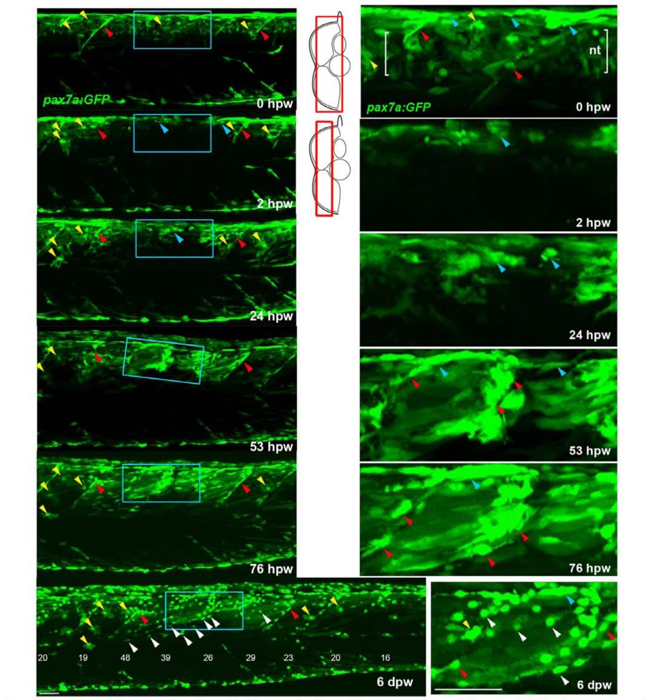
Fig. S7
Genetically marked pax7a-expressing cells contribute to regeneration. Tg(pax7a:GFP):pfe/pfe zebrafish larva was wounded at 4 dpf in the epaxial region of somites 16-19 and repeatedly confocally scanned live, embedded in agarose. Maximum intensity projections of selected regions of the stack are shown in lateral view, anterior to left, dorsal up. Only top panel includes neural tube levels (red boxes in schematics). Blue boxed regions, magnified at right, show GFP+ cells near dorsal epaxial edge in central somite 16 at 1 dpw yielding thin muscle fibres by 2 dpw (blue arrowheads). Prior to wounding (top panels), GFP is present in cells of neural tube (nt. between brackets), dorsal and vertical somite borders (blue and red arrowheads, respectively) and rare xanthophores (yellow arrowhead). In contrast to the larva in panel S6A, GFP recovers more rapidly in central wounded somites (blue box) that had GFP+ cells spreading from dorsal epaxial edge at 24 hpw (blue arrowheads). pax7a:GFP+ cells began to re-accumulate at the dorsal edge by 1 dpw and fibre formation was well underway by 2 dpw. As nascent fibres matured and increased in volume, their GFP fluorescent intensity declined, while small myogenic cel ls remained strongly labelled. In contrast to the larva in panel S6A, GFP recovers more rap idly in central wounded somites (blue box) that had GFP+ cells spreading from dorsal epaxial edge at 24 hpw (blue arrowheads). At 6 dpw, when wound repair was advanced, numerous small round GFP+ cells (white arrowheads) appear in all somites overlying the myotome, in the dermomyotome location. These cells were more numerous in wounded somites than in adjacent unwounded somites, and concentrated at the posterior somite border. In summary, pax7a-expressing myogenic cells contribute to muscle repair and a sub-population of such cells remains undifferentiated after myotome recovery. Bar = 50 µm .