
|
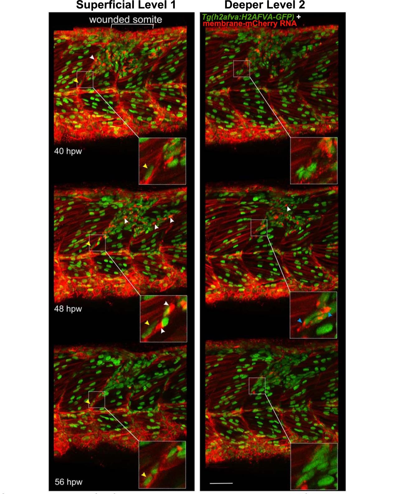
Fig. S5
Cells with specialised morphology at borders of wounded somites. Time-lapse of short stacks of lateral confocal sections from larva in Movie 81 , anterior to left. Wounded region is indicated (white bracket). Level 1 is about 30 µm more superficial than Level 2 revealing the extent of damage. Boxed insets show vertical myoseptal (VMZ) region. Some cells aligned with the VMZ (yellow arrowheads) move little between time-points. Other cells aligned at the VMZ had a small oval nucleus and a bright ′cap′ of membrane mCherry (white arrowheads) and appeared at the lesion site between 32-40 hpw, but could not be identified at the VMZ at subsequent time-points, suggesting rapid migration or change of appearance. At 48 hpw, such ′capped′ cells were dispersed in the central myotome and more numerous, but their number subsequently declined. Some cells at the VMZ appeared to have unusually elongated nuclei and bright bipolar cytoplasmic mCherry, suggesting that they might be dividing cells (blue arrowheads). The labelling of all cells and the low time resolution of the movie prevented observation of such events as MPC proliferation and fusion in real time. Bar = 50 µm.