
|
Fig. S4
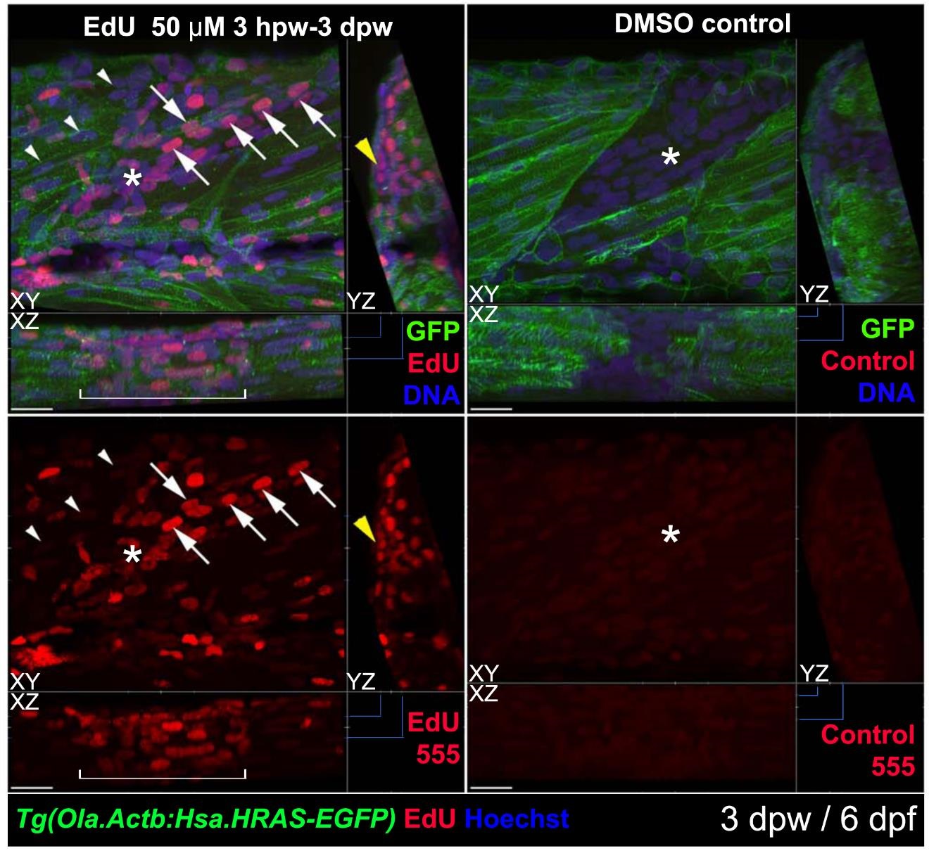
Most nuclei in wound sites have undergone S-phase. Tg(Ola.Actb:Hsa.HRAS-EGFP) larvae were wounded at 3 dpf in epaxial somite 517 and treated with EdU (left) or control vehicle (right) from 4 hpw until 3 dpw/6 dpf, followed by immunodetection of anli-GFP-Alexa488, ClickIT-555 for EdU and Hoechst for DNA. 3D confocal stacks were processed with Imaris to make short orthogonal projections from the planes indicated oy the blue markings. The wounded somite region (white bracket) contains abundant nuclei, most of which are EdU-marked. Many nuclei have the typical elongated form and alignment of fibre nudei (arrows). Other regions of the wound still contain nuclei with rounded morphology (asterisks). The transgene membrane GFP labels some regenerating cells weakly (right), for reasons that are unclear. Adjacent unwounded somites contain numerous nuclei unmarked by EdU (arrowheads) and some EdU-marked nuclei, possibly reflecting normal growth. Note the abundant EdU label in superficial cells reminiscent of dermomyotome (yellow arrowhead). 16/20 EdU-treated individuals showed all these features. Bars = 20 µm.