
|
Fig. S2
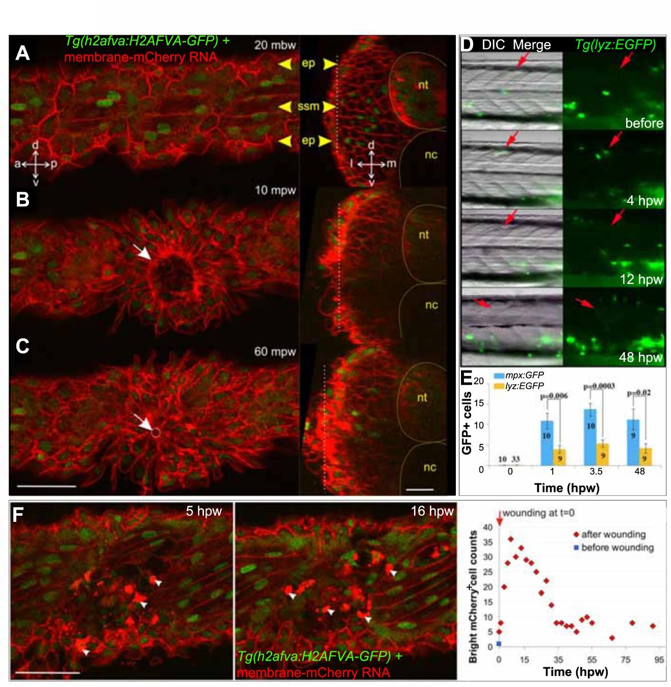
Epidermal and leukocyte movements during wound closure. A-C. Rapid epidermal wound closure in a 2.5 dpf membrane-mCherry RNA-injected Tg(h2afva:H2AFVA-GFP) larva in optical parasagittal section (left), taken at the level of the white dotted line in the transverse sections (right). A. Before wounding, polygonal epidermal cells (ep) overlie superficial slow muscle cells (ssm). B. At 10 mpw, an epidermal ring already formed (white arrow) and was almost closed at 60 mpw (C, white circle and arrow). Transverse sections show that the epidermal region above the wound thickens oy some 10-15 µm. D. Tg(lyz:EGFP) fish were wounded around s12 at 2.5 dpf (red arrows) and imaged in lateral view with dorsal up and anterior to left. GFP+ cells accumulated at wound site 4 hpw. E. Tg(mpx:GFP) and Tg(lyz:EGFP) were wounded in two to four somites between epaxial s5-12 at 2.5 dpf. GFP+ cells were counted at the wound site of the number of fish indicated. F. Change in numher of hright mCherry cells (arrowheads) with time in membrane-mCherry RNA-injected Tg(h2afva:H2AFVA-GFP) larva wounded at 2.5 dpf. a - anterior, d - dorsal. 1- lateral. m - medial, nc - notochord, nt - neural tube, p - posterior. v - ventral. Bars = 50 µm.