
|
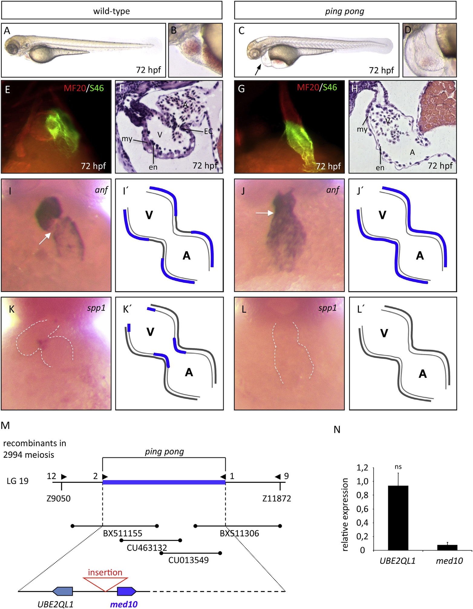
Fig. 1
Effects of the png mutation on heart development. (A-D) Wild-type (wt) (A) and png mutant (C) embryos and close-ups of the wt (B) and png (D) hearts at 72 hpf. (E, G) MF20 and S46 immunostainings reveal that chamber specification and differentiation is not altered in png mutants (E). (F, H) H&E staining of sections of wt (F) and png (H) hearts at 72 hpf. png mutant heart chambers are not separated by an AV ring and no endocardial cushions (EC) develop at the AVC, whereas png myocardial (my) and endocardial (en) cell layers are clearly defined. (I, J) anf is misexpressed in AV myocardial cells of png hearts (I), whereas anf is absent from wt AV myocardium (J) (AVC, white arrow). (K, L) Spp1 is expressed in AVC and OFT endocardium of wt embryos at 72 hpf (K), whereas spp1 expression is absent from the AVC of png hearts (L) (Schematic descriptions: I′, J′, K′, L′). (M) Integrated genetic and physical map of the png locus on chromosome 19. The png promoter mutation (1193bp insertion) is indicated (red). (N) The png mutation leads to severely diminished med10 transcription (relative expression 0.074 ± 0.039, n = 7, P < 0.001), whereas mRNA levels of UBE2QL1 are unaltered (relative expression 0.93 ± 0.18, n = 3, ns).