Fig. 1
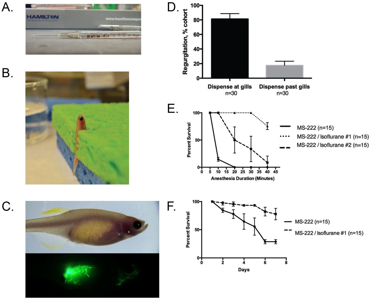
Technical optimization of oral gavage technique. (A) The gavage apparatus was constructed from a 10µl luer-tip Hamilton syringe with a 22G needle and 22G soft-tip catheter tubing. (B) A representative zebrafish was anesthetized using a MS-222/isoflurane combination anesthetic and immobilized vertically in a damp sponge with a slit. (C) A representative zebrafish was gavaged with 3µl fluorescein isothiocyanate-dextran (FITC-dextran) to visualize potential regurgitation via fluorescence microscopy. (D) Zebrafish were gavaged with Phenol Red solution to visualize potential regurgitation. Data represented as mean±s.d. of two replicates. (E) Survival curve for extended exposure to anesthetic solutions. Data represented as mean±s.d. of three replicates. (F) Survival curve for long-term daily exposure to anesthetic solution. Data represented as mean±s.d. of three replicates.

