
|
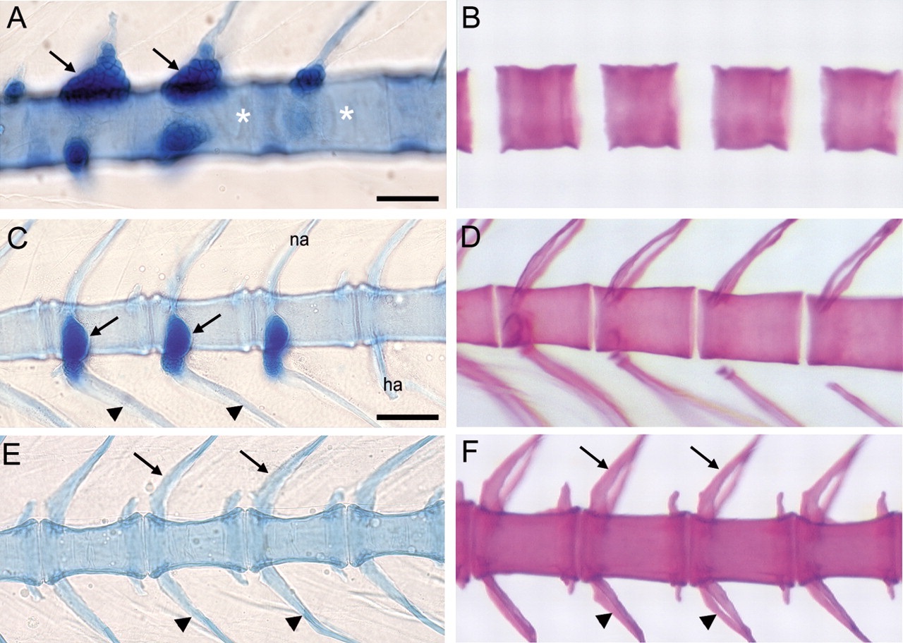
Fig. 1
Cartilage and bone formation in the zebrafish vertebral column. (A,C,E) Alcian green staining for cartilage. (B,D,F) Alizarin red staining for bone. (A) At 9 dpf cartilage is only found in the Weberian apparatus (arrows), a specialisation of the four most anterior vertebrae. The Weberian apparatus is described as arising from V1-5 by Morin-Kensicki et al. (Morin-Kensicki et al., 2002), although V1 and V2 give the appearance of a single vertebra in our experiments (see also Coburn and Futey, 1996). Staining is not seen in the centra (asterisks). The weak staining in the bulk of the centra is non-specific, acellular background staining, indicating an absence of chondrocytes and therefore cartilage in these regions. (B) Despite the absence of cartilage, bone is seen in cervical centra at 9 dpf. (C) Cartilage is only seen in the rib heads (arrows) and not in the centra, neural (na) or haemal (ha) arches, or distal ribs (arrowheads) at 17 dpf. (D) Ossified centra are present along the trunk by 17 dpf. (E,F) By 23 dpf, neural (arrows) and haemal (arrowheads) arches have also formed in the tail. Centra and arches ossify without forming cartilage. Scale bars: A,B,~ 50 µm; C-F, ~100 µm.