
|
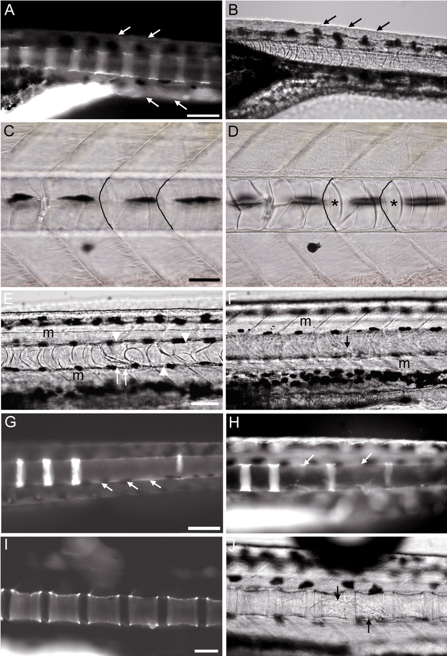
Fig. 4
Ablation of notochord cells prevents formation of centra. (A) Unablated embryo (12 dpf) stained with quercetin. Centra appear as stripes along the anterior-posterior axis. (B) DIC image of embryo in A; the anterior boundary of each centrum is level with each myoseptal border (arrows mark the myoseptal border cleft, anterior to left; extrapolation of the arrow vectors in A and B shows the alignment of the V-shaped myoseptal borders with the anterior edges of the developing centra). (C,D) Bright field images of a 4-dpf embryo, showing appearance of notochord cells prior to targeting with the laser. Cross hairs were aligned on the myoseptal borders at the focal plane of muscle segments (C), and the focus was then adjusted to the plane of notochord cells (D). Asterisks denote single notochord cells spanning the diameter of the notochord; these have a typically cylindrical appearance, and would be selected for ablation. The position of two myoseptal borders at the dorsoventral level of the notochord is indicated by dark lines. (E-H) Ablation of notochord cells before centrum formation (at 4 dpf) preventing subsequent development of centra. Cells were targeted at several positions along the anterior-posterior axis, each level with the myoseptal border (16 embryos). (E) DIC image of an embryo after notochord ablation showing the ablation site at 10 dpf (arrows); adjacent notochord cells have altered shape to fill the site (arrowheads). (F) Embryo in E at a focal plane above the notochord showing no damage in adjacent muscle (m); arrow indicates ablation site. (G) Targeting of three notochord cells from consecutive segments prevents development of three centra (arrows); quercetin labelling at 12 dpf. (H) Targeting of notochord cells from alternate segments prevents development of alternate centra (arrows); quercetin labelling at 12 dpf. (I,J) Ablation of notochord cells at 4 dpf in the centre of prospective centra does not affect their development (8 embryos). (I) Quercetin labelling at 20 dpf after ablation at 4 dpf. (J) DIC image of embryo in I, arrows indicate ablation sites. Scale bars: A,B, ~100 µm; C,D, ~50 µm; E,F, ~100µ m; G,H, ~100 µm; I,J, ~100 µm.