
Fig. 1
- ID
- ZDB-IMAGE-160823-11
- Genes
- Antibodies
- Source
- Figures for de Preux Charles et al., 2016
|
Fig. 1
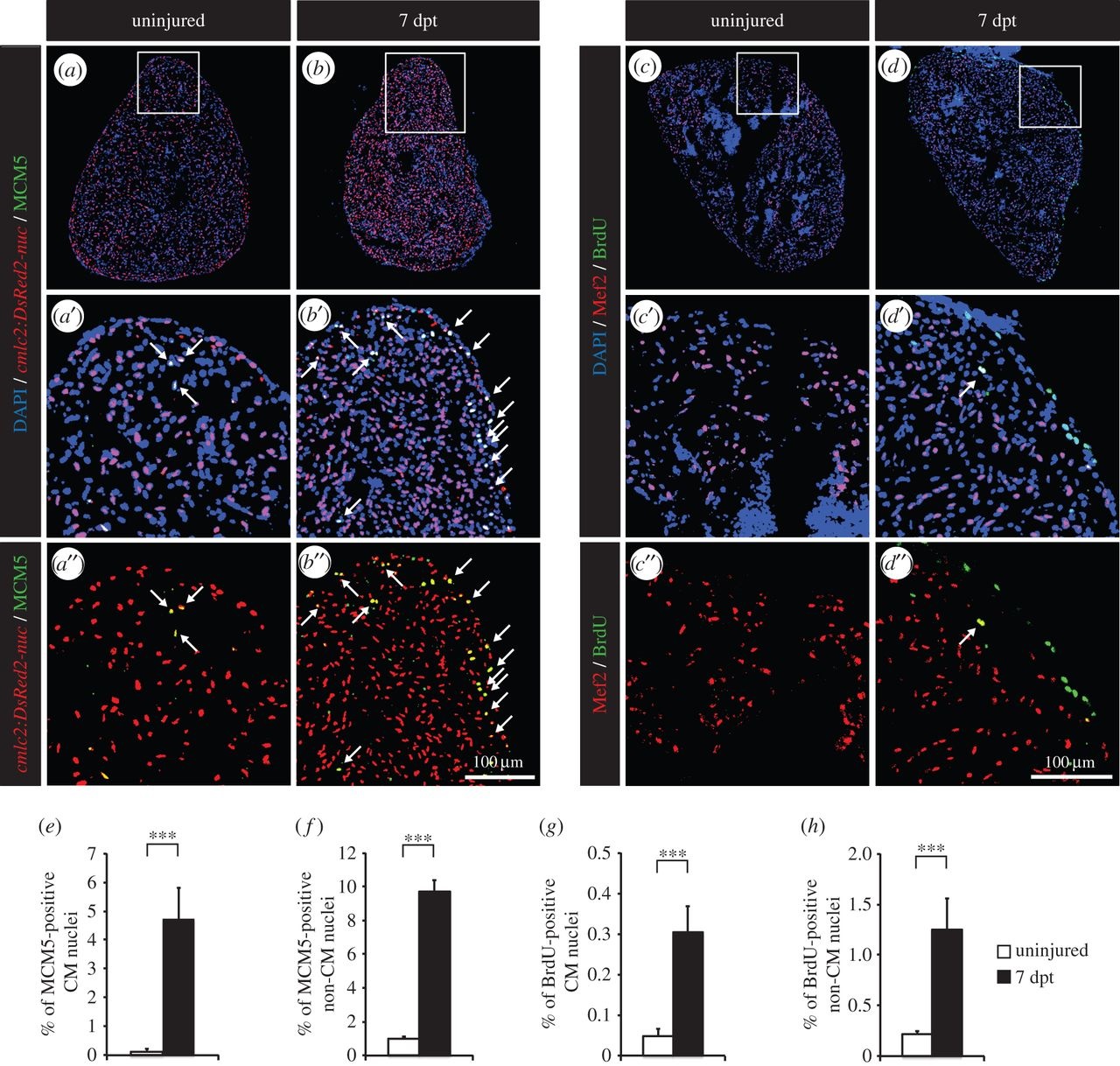
Thoracotomy triggers cell proliferation in the intact zebrafish heart. (a,b) Representative sections of hearts of transgenic fish cmlc2:DsRed2-nuc (red), which demarcate cardiomyocyte (CM) nuclei, labelled with the G1/S-phase marker MCM5 (green). (c,d) Representative sections of hearts after one week of BrdU (green) treatment. Mef2 staining (red) was performed to differentiate CM nuclei from non-CM nuclei. (a′,b′,c′,d′,a′′,b′′,c′′,d′′) Higher magnifications of the framed area shown in the images that are labelled with the same letter without the prime symbol. The same rule applies to all the subsequent figures. Arrows indicate double-positive nuclei. (e-h) Quantification of MCM5- and BrdU-positive CM and non-CM nuclei. (n ≥ 4 hearts; ≥2 sections per heart; ***p < 0.001).