
|
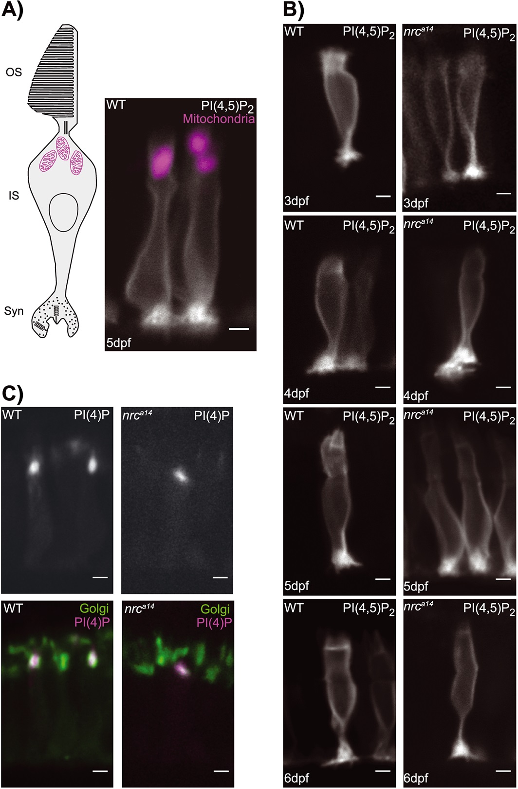
Fig. 6
PI(4,5)P2 and PI(4)P distributions are not altered in nrca14 cones. A and B: The PI(4,5)P2 probe GFP-PLCδ-PH was expressed in photoreceptors using the crx promoter. A: In wild type (WT) cells at 5 days post-fertilization (dpf), this probe localized to the plasma membrane and concentrated at synapses, but did not extend above the mitochondria (magenta) into the outer segment (OS). C: The PLC´-PH probe showed the same distribution in nrca14 photoreceptors from 3 to 6 dpf. PI(4)P was visualized with the probe RFP-FAPP1-PH. In both WT and nrca14 photoreceptors at 5 dpf, the RFP-FAPP1-PH probe (magenta) overlapped with the medial Golgi marker Man2a-GFP (green). There was no difference in the subcellular distribution of PI(4)P between WT and nrca14 photoreceptors. Scale bar = 2 µm in all images. IS, inner segment.