
|
Fig. 2
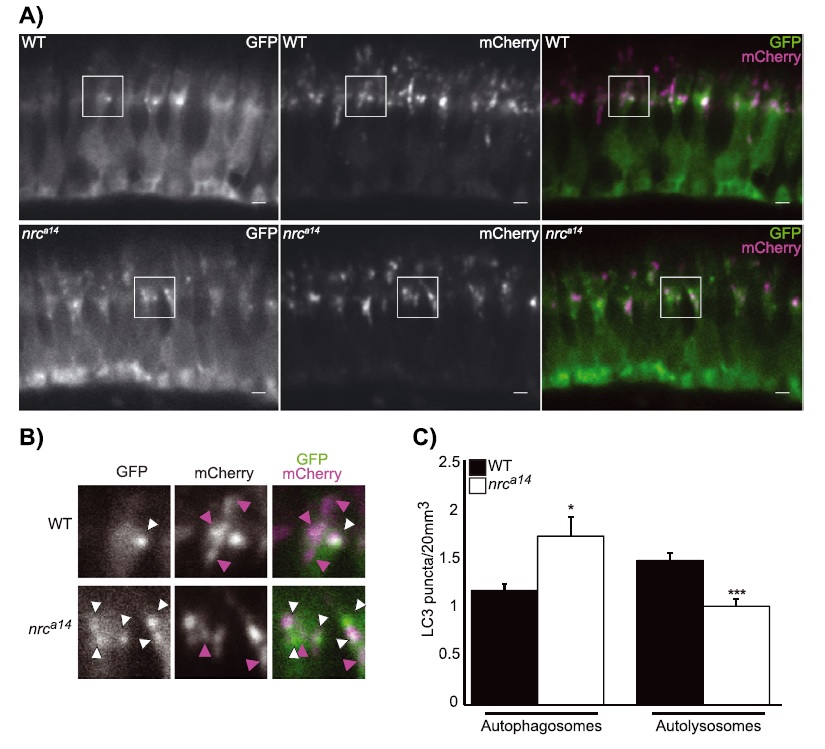
Maturation of autophagosomes is blocked in nrca14 cones. Expression of the tandem mCherry-GFP-LC3 probe in cones of wild type (WT) and nrca14 5 days post-fertilization larvae. A: In WT photoreceptors, autolysosomes (magenta-only puncta) were visible (top panel). A: Fewer magenta-only puncta were visible in nrca14 cones (bottom panel), indicating that the LC3 positive autophagosomes had not fused with an acidic compartment. B: Shows enlargement of areas shown in boxes in A. White arrow heads indicate GFP+, mCherry+ autophagosomes, magenta arrowheads point to GFP-, mCherry+ autolysosomes. Scale bar = 2 µm in A. Graph in C shows average number of autophagosomes (GFP+, mCherry+) and autolysosomes (GFP-, mCherry+) per 20 µm3. Error bars are SEM and n = 7 WT larvae and six nrca14 larvae. (***p-value < 0.001, *p-value < 0.05 respectively as assessed by Mann-Whitney test).