
Image
|
Figure Caption
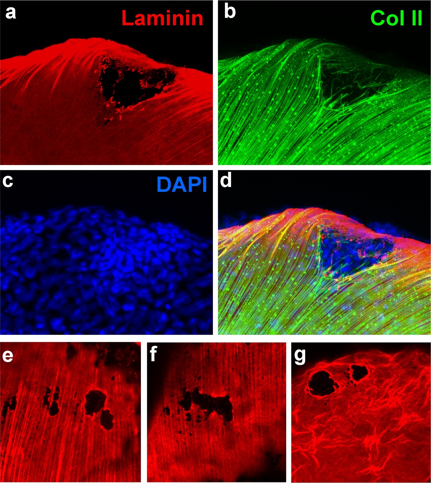
Fig. 2 S1
psoriasis mutants display local degradations of the skin basement membrane and of underlying collagenous actinotrichia of the dermis.
(a-d) IF of laminin (red; a) and type II collagen (green; b) in a whole mount 58 hpf psoriasis mutant fin fold, counterstained with DAPI (blue; c). The basement membrane is disintegrated (a) and actinotrichia (b) are disassembled below an epidermal aggregate (d; merged channels). (e-g) IFs of laminin (red) show examples of holes in the basement membrane in different 58 hpf psoriasis mutants.
Acknowledgments
This image is the copyrighted work of the attributed author or publisher, and
ZFIN has permission only to display this image to its users.
Additional permissions should be obtained from the applicable author or publisher of the image.
Full text @ Elife