
|
Fig. 5
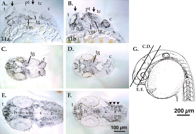
The sox11a and sox11b genes are expressed in overlapping and unique regions of the embryonic brain. A,B: Cells in the dorsal telencephalon, pretectum and tectum express sox11b (B), but not sox11a (A, arrows). Brain regions with similar sox11 gene expression domains include the telencephalon, diencephalon, ventral midbrain, and tegmentum. Ventral midbrain expression of sox11b is weaker than sox11a expression. The arrows indicate domains of differential gene expression in the dorsal telencephalon (left arrow in A and B) and the tectum (right arrows). C,D: sox11a and sox11b are expressed in the tegmentum and the cerebellum. The plane of sectioning is slightly more dorsal in C. E,F: In the ventral brain, ventral mesencephalic cells express sox11a and sox11b. However, sox11a expression is stronger posterior to the hypothalamus (posterior ventral diencephalon) and ventral midbrain than sox11b. Section 5F is a deep horizontal section, and median cells lacking sox expression in the cerebellum are in the floor plate region. A,B sagittal sections; C-F horizontal sections, as indicated in G. t, telencephalon; h, hypothalamus; tg, tegmentum; pt, pretectum; tc, tectum; vm, ventral mesencephalon; c, cerebellum; r, rhombencephalon. Scale bars = 100 µm in A-F, 200 µm in G.