
|
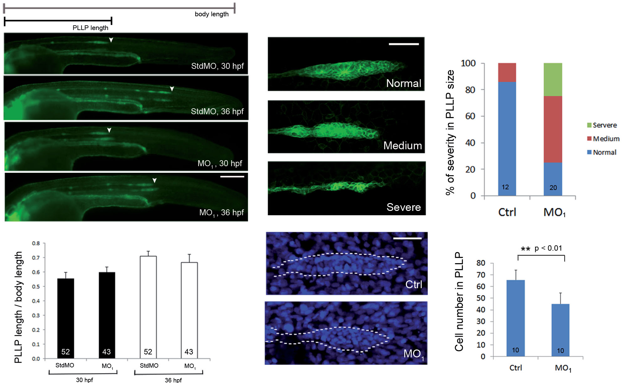
Fig. 8
Loss of calnexin reduced primordium size but did not affect migration of the posterior lateral line primordium (PLLP). (A-D) Transgenic claudin b:gfp embryos were treated with 5 ng std MO and with 3.75 ng of the calnexin MO1 and imaged at 30 and 36 hpf. Embryos were observed and photographed under epifluorescence microscopy. Scale bar: 200 µm. (E) Ratios of the migration path of the PLLP to body length at 30 and 36 hpf are shown. (F-K) Transgenic claudin b:gfp embryos were treated without or with MO1, cultured until 32 hpf, stained with DAPI and examined under confocal microscopy for GFP (excitation: 455-490 nm; emission 500-540 nm) and DAPI signal (excitation: 350 nm; emission 470 nm), respectively. Representative confocal photographs of different severity of PLLP defects are shown in (F-H) and the percentages of embryos with different severities are shown in (I). Photographs of control embryos (J) and MO1 morphants (K) stained with DAPI are shown. (L) The average cell numbers in PLLPs are shown. * *Indicates that the value significantly differs from that of control embryos (p < 0.001).Scale bar: 50 +m. Photographs are shown in lateral view with the anterior to the left and posterior to the right.