
|
Fig. 4
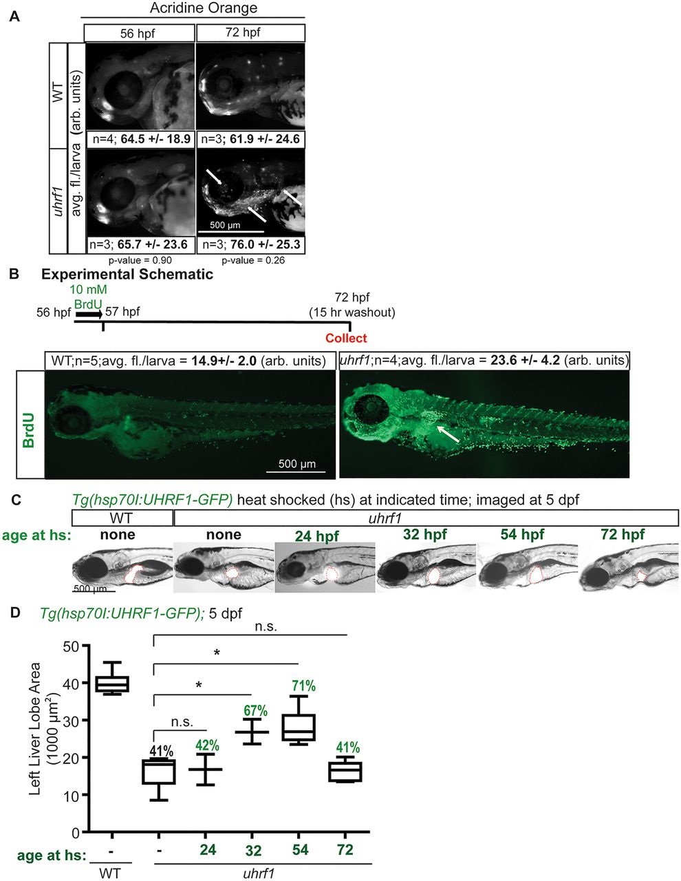
Cell cycle arrest is an acute response to the loss of uhrf1. (A) Acridine Orange staining of whole fish was imaged at 56 and 72hpf. Averaged intensity of fluorescence from the head, jaw and ear of three or four larvae (n) is indicated with the s.d. Arrows point to the regions with enhanced cell death in mutant larvae. (B) Whole fish were pulsed with BrdU at 56hpf for 1h and processed for BrdU immunofluorescence at 72hpf. Arrow points to intense labeling in the liver. (C) Tg(hsp70I:UHRF1-GFP; fabp10:ds-Red);uhrf1 and wild-type siblings were heat-shocked to induce expression of UHRF1 at the indicated times. The liver is outlined in red. (D) The left liver lobe area was quantified in three or four fish per time point. All fish contain the Tg(hsp70l:UHRF1-GFP) transgene. Heat shock had no effect on liver size in WT fish. Box plots are as described in Fig. 3E. *P<0.05 by Student′s t-test. The percent difference in the area of the left liver lobe of the mutants relative to wild-type is indicated.