
Image
|
Figure Caption
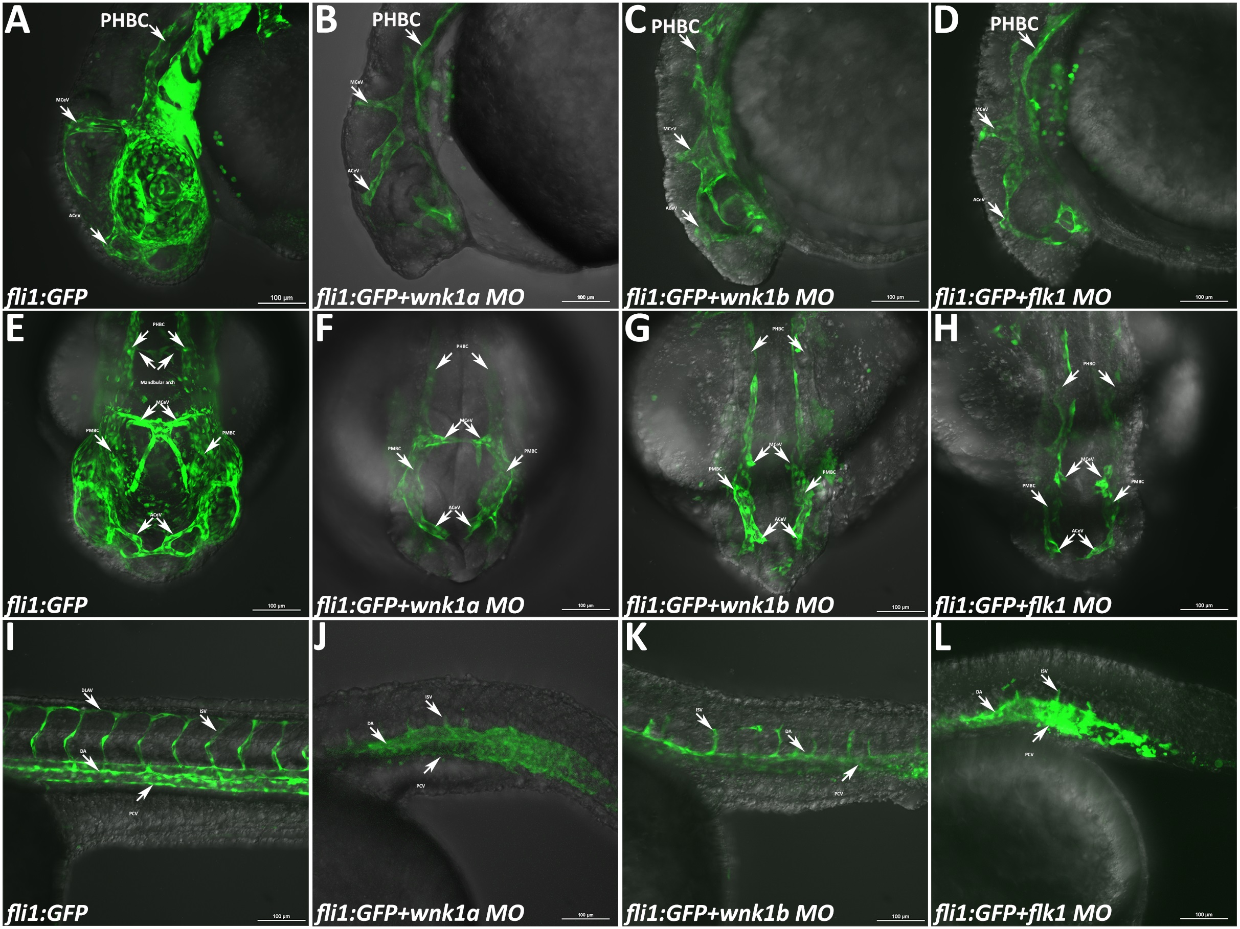
Fig. 4
Phenotype of Tg(fli1:GFP) embryos injected with various morpholinos and imaged with a confocal microscope.
(A-D) Lateral views of the heads of uninjected control embryos and wnk1a, wnk1b and flk1 morphants at 33 hpf. (E-H) Frontal views of the heads of uninjected control embryos and wnk1a, wnk1b and flk1 morphants at 33 hpf. (I-L) Lateral views of the trunk in uninjected control embryos and wnk1a, wnk1b and flk1 morphants at 33 hpf. Important vessels are indicated with arrows and labeled, with the full name given in the text. Scale bar: 100 µm.
Figure Data
Acknowledgments
This image is the copyrighted work of the attributed author or publisher, and
ZFIN has permission only to display this image to its users.
Additional permissions should be obtained from the applicable author or publisher of the image.
Full text @ PLoS One