
|
Fig. 1
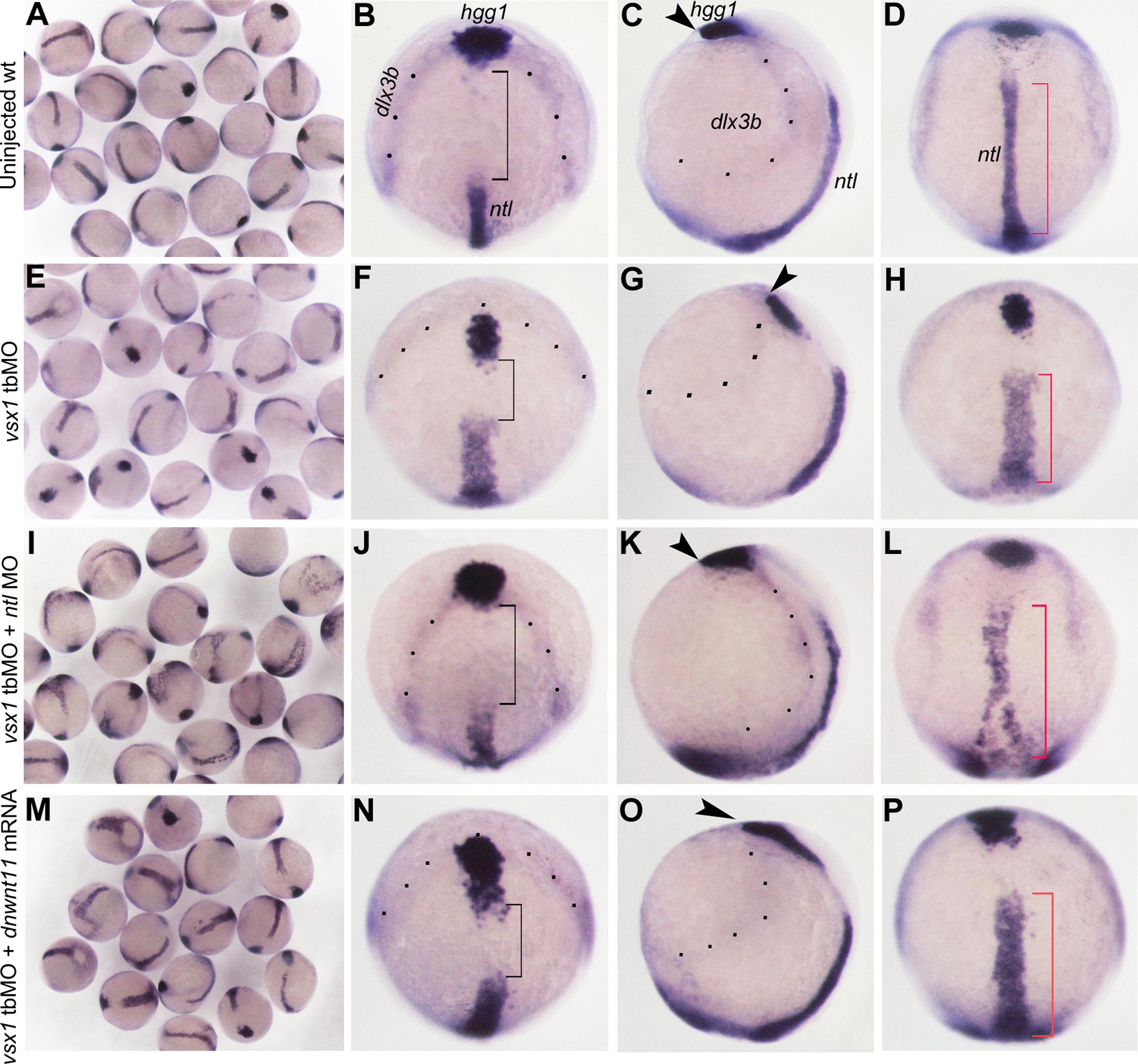
Comparison of spatial relationship patterns of PME, axial mesoderm, neural plate and epidermis in different types of embryos at bud stage. The neural plate and epidermis boundary is marked by dlx3b, PME is marked by hgg1 and axial mesoderm is marked by ntl. (B, F, J, N) Dorsal-animal views with anterior towards the top. (C, G, K, O) Lateral views with animal towards the top. (D, H, L, P) Dorsal views with animal towards the top. The injected reagents are indicated at the left of each column. The border between neural plate and epidermis is emphasized by black dots. Black brackets indicate the distance between anterior prechordal plate and posterior notochord. Red brackets indicate the length of notochord. Arrowheads indicate the anterior position of PME. Note that in vsx1 tbMO and ntl MO coinjected embryos, the anterior migration of hgg1 expression domain and the extension of ntl expression domain are recovered, but the lateral expansion of ntl expression domain is still the same as that in vsx1 morphants.
Reprinted from Developmental Biology, 394(2), Xu, X., He, Y., Sun, L., Ma, S., Luo, C., Maternal Vsx1 plays an essential role in regulating prechordal mesendoderm and forebrain formation in zebrafish, 264-76, Copyright (2014) with permission from Elsevier. Full text @ Dev. Biol.