
|
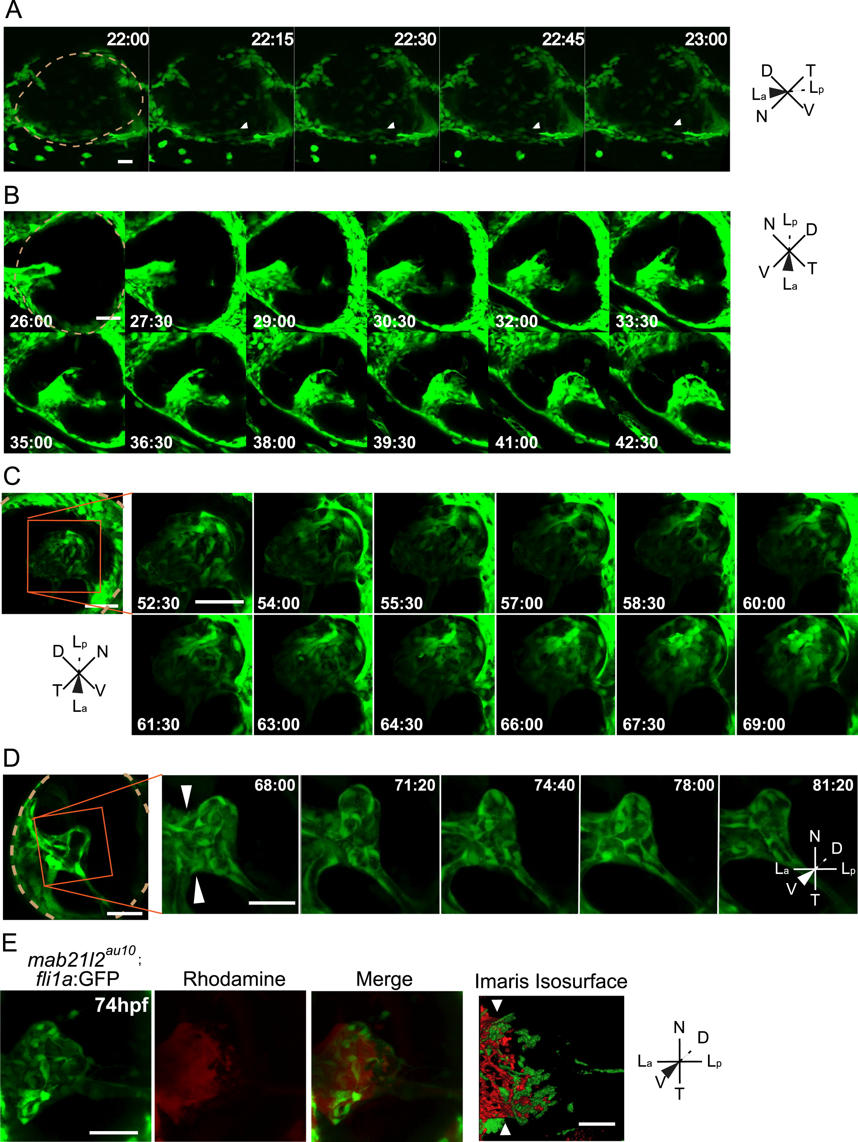
Fig. 8
The lens is required for Stages II and III hyaloid maturation and maintenance. All images are stills from time-lapse movies from severe mab21l2au10; fli1a:GFP mutants. (A) Hyaloid precursor cell recruitment is delayed by ~2 h in mab21l2au10 mutants but recruitment is otherwise normal. (B) Hyaloid loop formation occurs in the absence of a lens. (C) Stage II formation of a branched hyaloid network is disrupted in mab21l2au10 mutants. Hyaloid cells appear disorganized and dynamic, having not coalesced into obvious vessels. (D) The hyaloid in mab21l2au10 mutants still makes contact anteriorly with the annular vessel. (E) Microangiography demonstrates that at 3d pf, the mab21l2au10 mutant hyaloid is not enclosed anteriorly, and rhodamine (red) fills the retina. hh:mm. Beige dashed line in A, B and D indicate outlines of the retina. All scale bars=50 µm. D: Dorsal, V: Ventral, N: Nasal, T: Temporal, La: Lens anterior, Lp: Lens posterior.
Reprinted from Developmental Biology, 394(2), Hartsock, A., Lee, C., Arnold, V., Gross, J.M., In vivo analysis of Hyaloid vasculature morphogenesis in zebrafish: A role for the lens in maturation and maintenance of the Hyaloid, 327-39, Copyright (2014) with permission from Elsevier. Full text @ Dev. Biol.