
|
Fig. S3
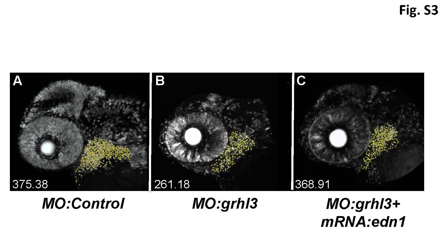
Pharyngeal arch regions used for IMARIS cell-proliferation quantitation. Representative confocal images of EdU-labelled controls (A), grhl3-morphants (B) and edn1-rescued grhl3-morphants (C), together with automated cell counts per image. Embryos were imaged with the Olympus Inverted FV1000, and pharyngeal arch (PA) cell counts were completed using IMARIS V7.6. Here, BA1 and 2 were selected (in the x, y and z axis) using the surface tool to both calculate arch volume, and create a new channel using the mask selection tool. Cell counts were completed on this new channel using the spots tool with estimated diameter set to 5µm, background subtraction on, sphere detection on, and IMARIS quality threshold minimum set to 45-70 (quality threshold maximum was set to maximum).
Reprinted from Mechanisms of Development, 133, Dworkin, S., Simkin, J., Darido, C., Partridge, D.D., Georgy, S.R., Caddy, J., Wilanowski, T., Lieschke, G.J., Doggett, K., Heath, J.K., Jane, S.M., Grainyhead-like 3 regulation of endothelin-1 in the pharyngeal endoderm is critical for growth and development of the craniofacial skeleton, 77-90, Copyright (2014) with permission from Elsevier. Full text @ Mech. Dev.