
|
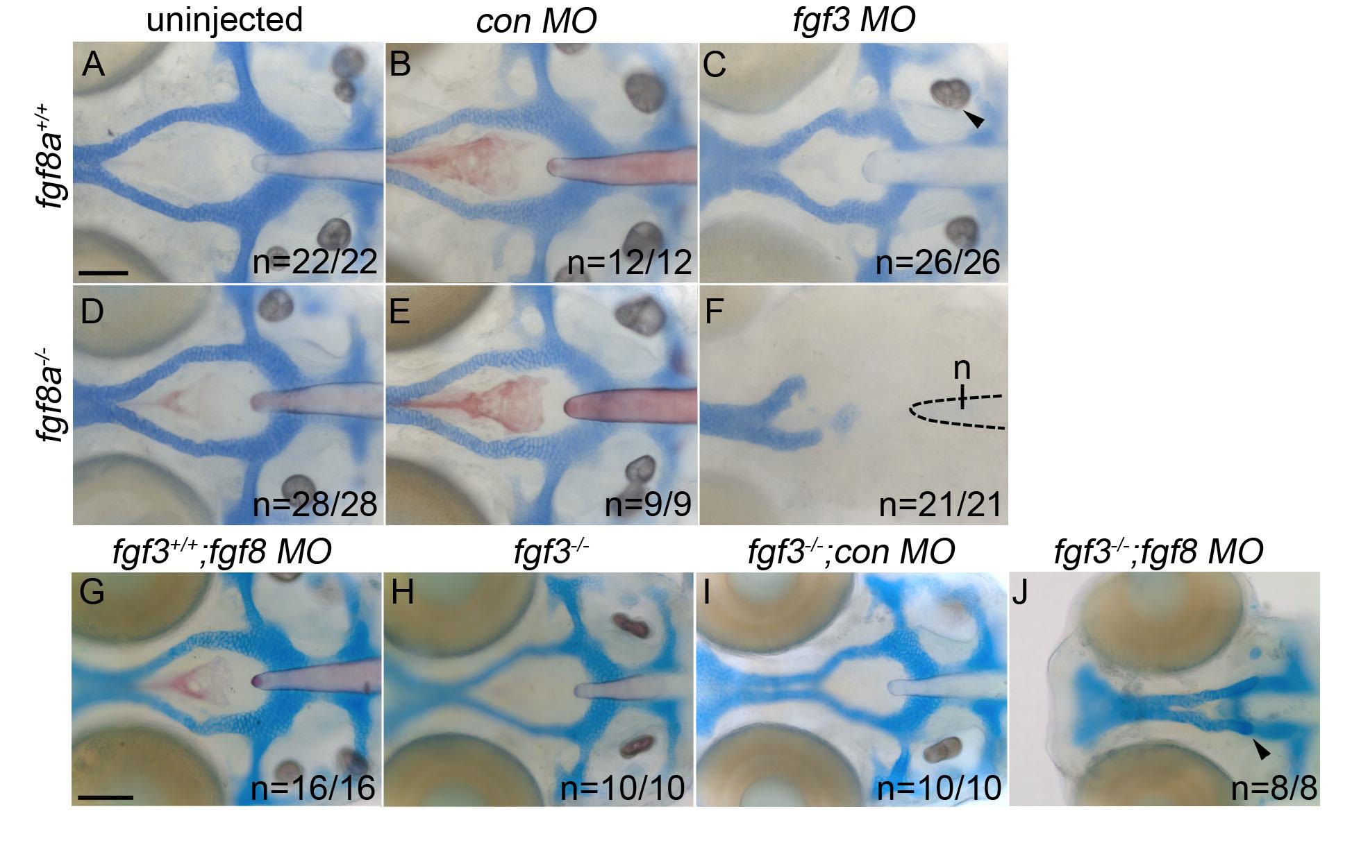
Fig. S1
Loss of Fgf signaling causes postchordal neurocranial defects. (A-J) Wholemount zebrafish neurocrania at 5 days post fertilization, with the viscerocrania removed. Anterior is to the left. Wildtype embryos injected with (B) control or (C) fgf3-morpholinos develop normal posterior neurocranial structures compared to (A) uninjected controls. (D and E) Un-injected and control morpholino-injected fgf8a mutants display normal posterior neurocranial development compared to (A) wildtype controls, but (F) develop severe posterior neurocranial loss when injected with fgf3 morpholino. (G) Wildtype injected with fgf8 morpholino, (H) fgf3 mutants, and (I) control morpholino-injected fgf3 mutants display normal neurocranial development however, (J) fgf8 morpholino-injected fgf3 mutants display severe postchordal neurocranial scale bar=20 µm.
Reprinted from Developmental Biology, 415(2), McCarthy, N., Sidik, A., Bertrand, J.Y., Eberhart, J.K., An Fgf-Shh signaling hierarchy regulates early specification of the zebrafish skull, 261-77, Copyright (2016) with permission from Elsevier. Full text @ Dev. Biol.