
|
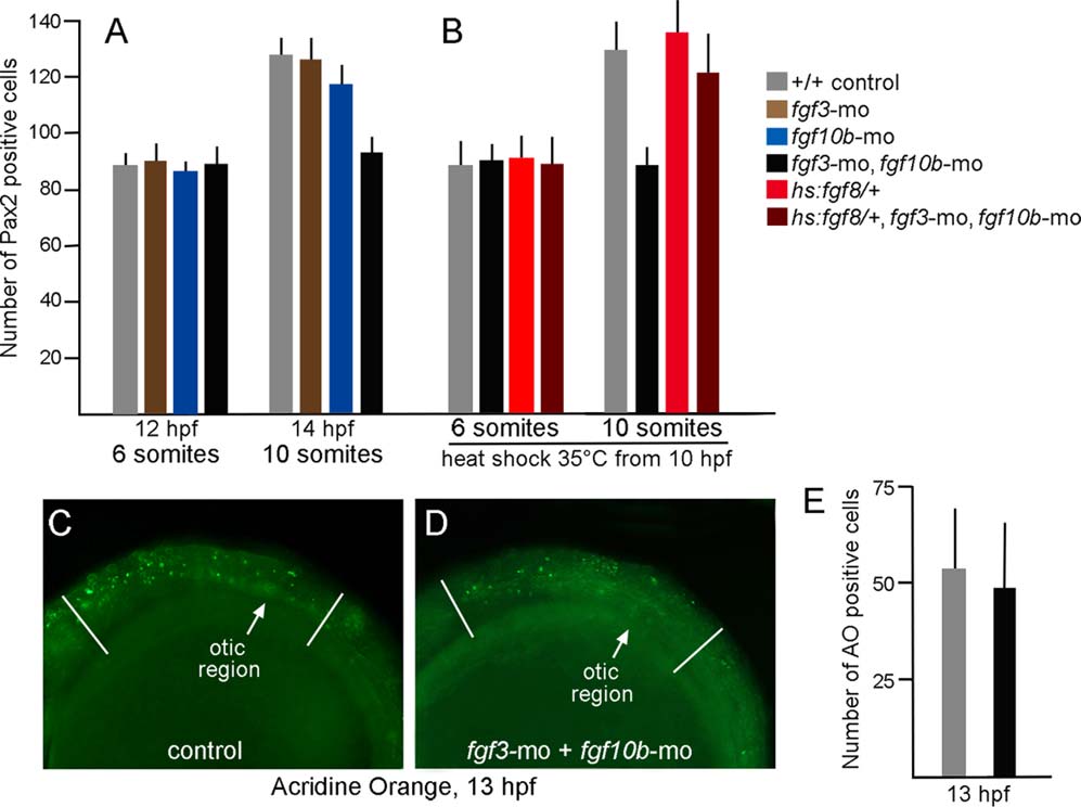
Fig. 4
Otic deficiencies in fgf3-fgf10b double morphants and rescue by timed misexpression of fgf8. A,B: The number of Pax2-expressing cells in the otic vesicle at 12 hpf and 14 hpf in embryos with various genotypes and gene knockdown, as indicated in the color key. Embryos in (B) were incubated at 35°C from 10 hpf until fixation at the 8- or 10-somite stage. Note that incubation at 35°C accelerates development such that these stages occur slightly earlier than normal. C,D: Comparison of the number of apoptotic cells stained with acridine orange (AO) in the head between the eye and first somite (marked by white lines), as seen from a lateral view in control embryos (C) or fgf3-fgf10b double morphants (D). E: Data show means and standard deviations of eight specimens each.