
|
Fig. 3
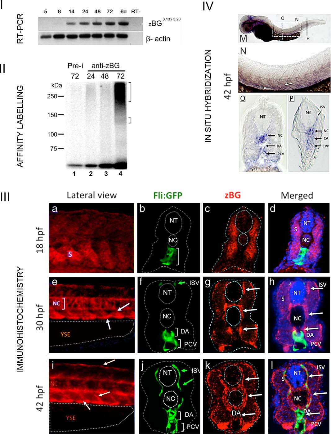
Expression of BG in zebrafish embryos. I: RT-PCR analysis of BG mRNA expression. Electrophoresis in agarose gel of samples corresponding to the RT-PCR products from T/W embryos RNA using the primers for zebrafish BG (see experimental procedures and Supporting Information Fig. S1). A single band of was obtained, corresponding to the expected 738 bp product of BG cDNA. This band was only detected at stages 14 hpf or later. Beta-actin was used as a loading control, and RT- indicates the control without reverse transcriptase enzyme. II: BG protein expression in zebrafish embryos. Electrophoresis in polyacrylamide gel- Samples correspond to total protein extracts that were subjected to TGF-β2 binding followed by immunoprecipitation with rabbit anti-zBG serum # 31, and crosslinking as described in Materials and Methods (see Supporting Information Fig. S1). From 24 to 72 hpf (lanes 2-4), the anti-BG serum detected a labeled smeary band centered around 250 kDa, as well as faint sharper band at ~120-130 kDa. Control was done with preimmune serum (Pre-i, lane1, corresponds to 72 h). III a-l: Immunohistochemistry (IHC) in TW embryos. Representative examples of embryos processed for IHC using a rabbit antiserum generated against zebrafish BG. a, e, and i: Lateral views of the trunk at the level of caudal limit of the YSE at the indicated stages. Red staining corresponds to BG expression. Anterior is to the left, dorsal to top. b-d, f-h, j-l: Transversal sections of Tg(fli:EGFP)y1 embryos at the level of the YSE at the indicated stages. Dotted lines were added to help identify the limits of the ectoderm, the neural tube and the notocord. b: At 18 hpf, GFP expression was found in ECs in the future DA and posterior Caudal Vein (PCV) (white bracket). c: In the same section high BG expression was localized in somites and low or absent in the notochord (NC). d: Merged image including DAPI staining of nuclei. e: Lateral views of the trunk of a 30 hpf embryo showing high expression of BG in the ventral aspect of somites, intersomitic boundaries (arrows) and NC (bracket). f: At 30 hpf, GFP expression was found in the DA and PCV and ISV. Note that in this section only the dorsal most tip of ISVs is seen (green arrow). g: In the same section, BG expression (white arrows) was high in cells at periphery of ventral somites, the NC and surrounding the neural tube. h: Merged image showing that sites of BG expression sites are adjacent to the DA, PCV, and ISV. i: Lateral views of the trunk of a 42 hpf embryo showed expression of BG similar to 30 hpf (white arrows), including now the dorsal aspect where the DLAV develops. j: At 42 hpf GFP expression indicated ECs of the DA, PCV, and ISV (green arrows). k: Note that BG expression was localized at higher levels (white arrows) in the NC and cells immediately adjacent to it, as well as the periphery of somites and neural tube. l: Merged image showing that sites of high BG expression are adjacent to GFP positive cells of the DA, PCV, and ISV. Abbreviations: Notochord (NC), Neural Tube (NT), ISV, Caudal Artery (CA), Posterior Caudal Vein (PCV), YSE, Somite (S)-. IV (m-p): “Whole-mount In situ hybridization” in TW embryos. Representative example of embryo processed for ISH using a riboprobe specific for zebrafish BG (nucleotides 1-861 of the cloned cDNA). m: Lateral view of a whole embryo at 42 hpf. Dotted rectangle indicates the magnified region shown in N. n: Lateral view at higher magnification of the indicated regions of embryo in M. BG expression was found in ventral somites and intersomitic boundaries. o: transverse cryostat section at the level of the YSE (indicated in M) showing expression of BG in the NC and somitic cells surrounding DA and PCV (black arrows). p: transverse cryostat section at the level of the CVP (indicated in M) showing expression of BG in the NC and somitic cells surrounding DA and CVP (black arrows) and ISV. Abbreviations: Notochord (NC), Neural Tube (NT), ISV, Caudal Artery (CA), Posterior Caudal Vein (PCV), YSE, Caudal Vein Plexus (CVP), and ISV.