
|
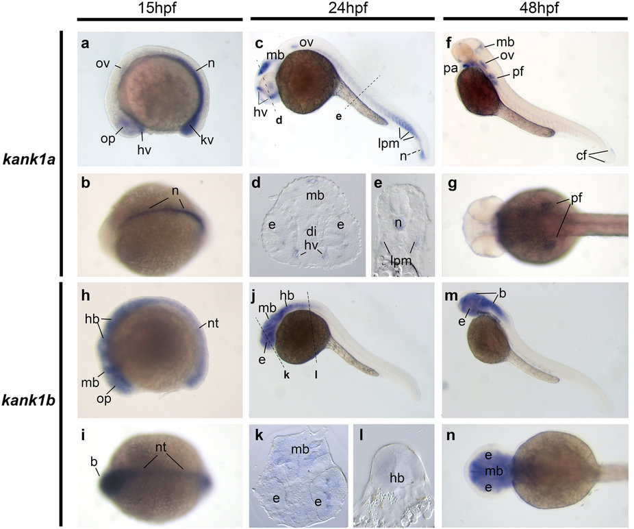
Fig. 3
Gene expression patterns of kank1a and kank1b in zebrafish embryos.
Whole-mount in situ hybridization of zebrafish embryos at stages 15 hpf (a,b,h and i), 24 hpf (c-e,j-l) and 48 hpf (f,g,m,n). Anterior is to the left in all the whole-mount images, and dorsal is to the top in all transverse sections. (a,b). Lateral and dorsal view of the expression of kank1a at 15 hpf. (c,f). Lateral view of kank1a expression at 24 hpf and 48 hpf, respectively. (h,i). Lateral and dorsal view of the expression of kank1b at 15 hpf. (j,m). Lateral view of kank1b expression at 24 hpf and 48 hpf, respectively. The dashed lines indicate the positions of sections. The letters below the dashed lines correspond to the panels. b, brain; cf, caudal fin bud; di, diencephalon; e, eye; hb, hindbrain; hv, head blood vessels; kv, Kupffer’s vesicle; lpm, lateral plate mesoderm; mb, midbrain; n, notochord; nt, neural tube; op, optic vesicle; ov, otic vesicle; pa, pharyngeal arch; pf, pectoral fin.