
Image
|
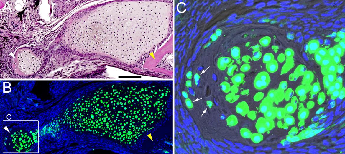
Figure Caption
Fig. S4
Contribution of col2a1aBAC:GFP-derived cells to bone. (A) H&E staining at 30 dpr shows remnant cartilage surrounded by bone (white arrowhead) which has a similar appearance to bone on the right (yellow arrowhead). (B) An adjacent section from this col2a1aBAC:GFP animal was processed for anti-GFP staining (green). Hoescht labels nuclei in blue. (C) Magnification shows that GFP+ cells are embedded in bone (arrows). A Normarski channel is included to show bone matrix. Scale bar = 100 microns.
Acknowledgments
This image is the copyrighted work of the attributed author or publisher, and
ZFIN has permission only to display this image to its users.
Additional permissions should be obtained from the applicable author or publisher of the image.
Full text @ Development