
Image
|
Figure Caption
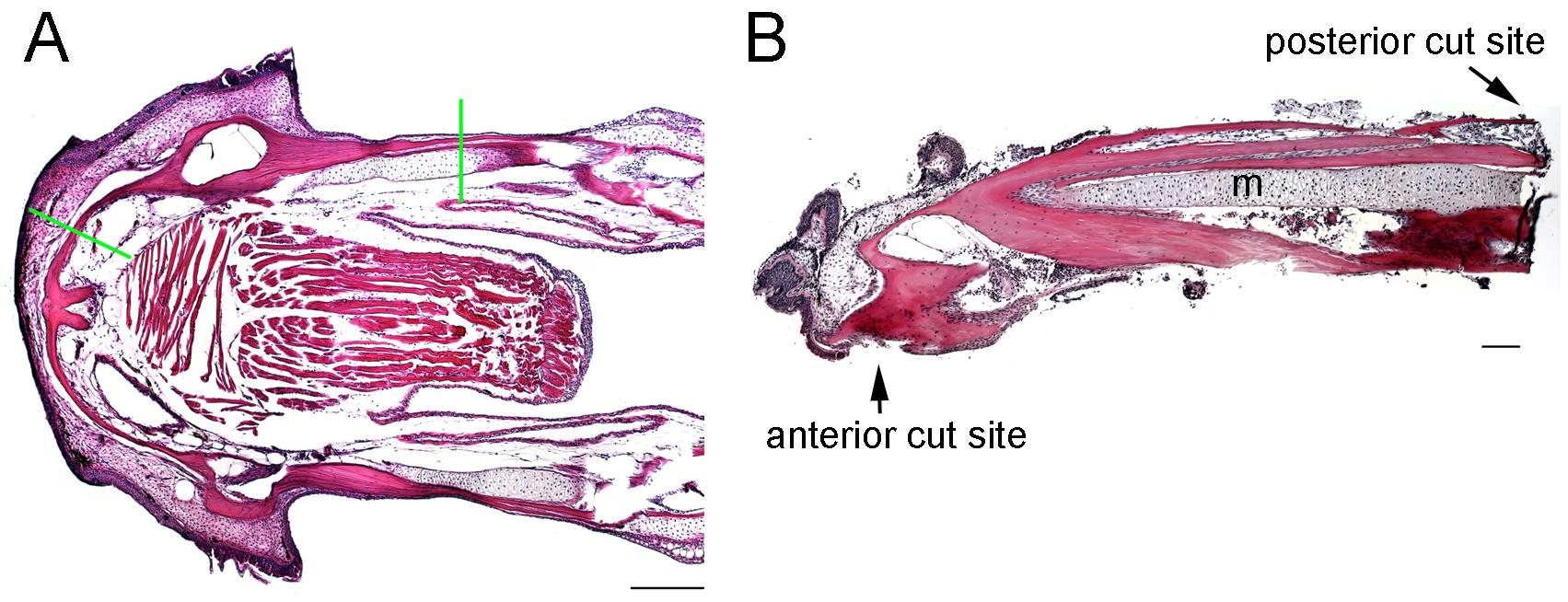
Fig. S1
Extent of lower jaw resections. (A) Coronal section of un-resected adult lower jaw stained with H&E. Green lines show where resection cuts are made. Anterior is to the left. (B) Histological section through the tissue that was removed showing the extent of bone removed and complete removal of the distal end of Meckel’s cartilage (m). Scale bars: A, 1 mm; B, 100 microns.
Acknowledgments
This image is the copyrighted work of the attributed author or publisher, and
ZFIN has permission only to display this image to its users.
Additional permissions should be obtained from the applicable author or publisher of the image.
Full text @ Development