
|
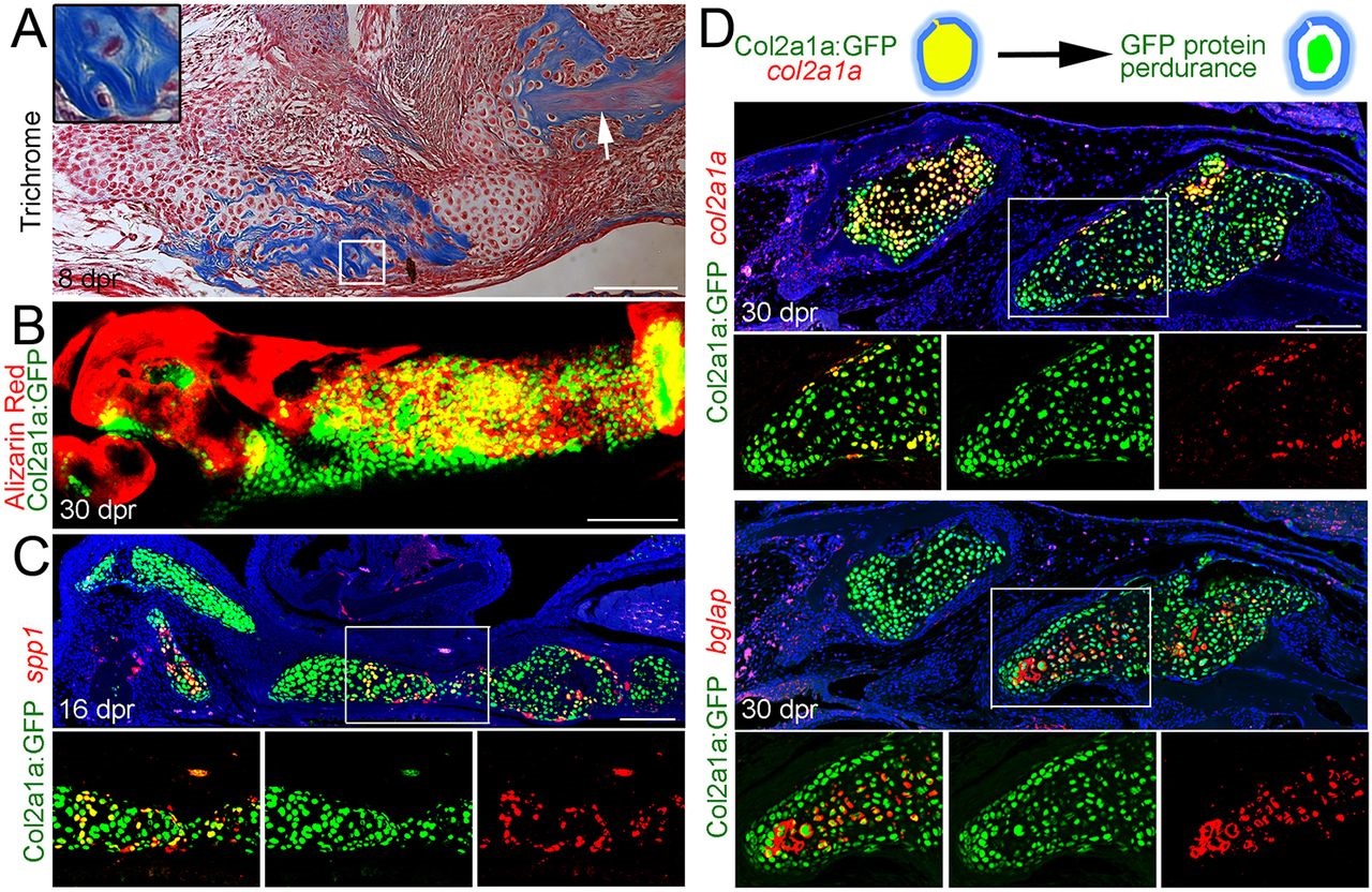
Fig. 3
Mineralization and osteoblastic maturation of repair chondrocytes. (A) Trichrome staining at 8dpr reveals the presence of collagen-rich osteoid-like material surrounding individual chondrocytes of the regenerating jaw. Inset shows a magnified view of several chondrocytes. Note the similar appearance of collagen staining (blue) between the unresected jawbone (arrow) and the mineralizing cartilage. (B) Alizarin Red staining of the regenerating jawbone in live adult zebrafish shows mineralization of col2a1aBAC:GFP-expressing chondrocytes. (C) Fluorescent in situ hybridization together with anti-GFP immunohistochemistry shows a subset of col2a1aBAC:GFP+ chondrocytes co-expressing the osteoblast marker spp1. (D) Schematic showing that GFP protein perdures in cells long after endogenous col2a1a mRNA ceases to be produced. In the fluorescent in situ images of regenerating jawbone at 30dpr, cells of the left callus still express col2a1a whereas cells of the right callus have largely ceased col2a1a expression and now express high levels of bglap (osteocalcin). Retention of GFP protein shows that bglap+ cells arise from cells that previously expressed col2a1aBAC:GFP. Areas in the white boxes are magnified and presented as merged and individual channels. Scale bars: 100µm.