
|
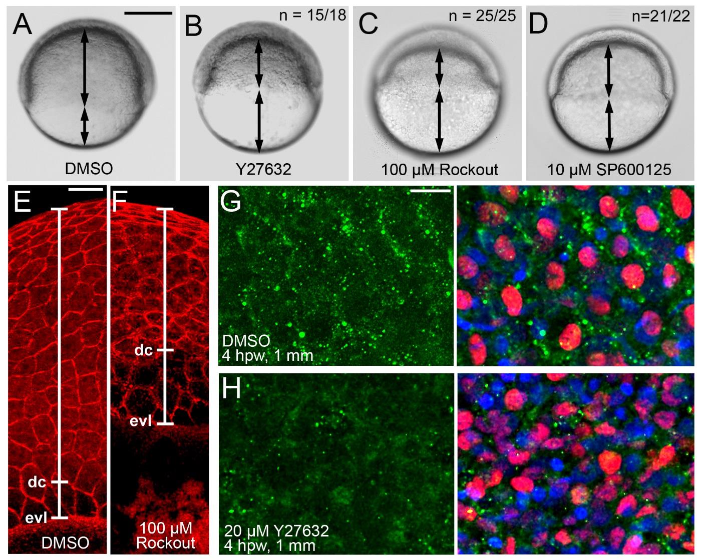
Fig. S8
Chemical inhibition of Rock or JNK leads to compromised epiboly movements in gastrulating embryos and to reduced p-myosin II levels in the epidermis around full-thickness wounds of adult zebrafish. (A-D) Live images of embryos at 7 hpw (70% epiboly stage for control; A). Embryos shown in (A,C,D) had been incubated in medium containing the indicated drugs at the indicated concentration, starting at 4 hpf; embryo shown in (B) had been microinjected with Y-27632 at the 1-cell stage (2 nl of 400 µM solution). Regions covered by deep cells and uncovered yolk are indicated by arrows. The outer enveloping layer (EVL) is not visible in these images. Rock and JNK inhibition lead to compromised deep cell epiboly. (E,F) Embryos as shown in (A,C), after phalloidin staining of actin cytoskeleton. In addition to deep cells, Rock inhibition also leads to delayed epiboly of the EVL, possibly pointing to an essential role of Rock during EVL cell flattening, which is a driver a EVL epiboly (Slanchev et al., 2009). However, deep cell (dc) epiboly is more severely compromised than EVL (evl) epiboly, indicated by the increased distance between the margins of the two tissues on the yolk (marked by white line). These differential and dissectable effects point to an additional role of Rock during radial intercalation, which is a major driving force of deep cell, but not EVL epiboly, consistent with results obtained in former mutant analyses (Slanchev et al., 2009). (G,H) Confocal images of epidermis at 1 mm distance from full-thickness wound of adults, 4 hpw, immunolabeled for phosphorylated non-muscle myosin (green) and p63 (red), and counterstained with DAPI (blue). Left panels show pMyosin channel, right panels merged images. p63+ keratinocytes of the DMSO-treated control fish display stronger pMyosin labelling particularly in cortical regions (G), while labelling in the Y27632-treated fish is much reduced and less localized (H). The punctate localization in (G) is in line with the reported punctate actin contractions taking place during morphogenetic movements in Xenopus embryos (Kim and Davidson, 2010). Higher nuclei density in (H) is a reflection of the compromised thinning of the epidermis. Scale bars: A-D: 200 µm; E,F: 50 µm; G,H: 10 µm.