
|
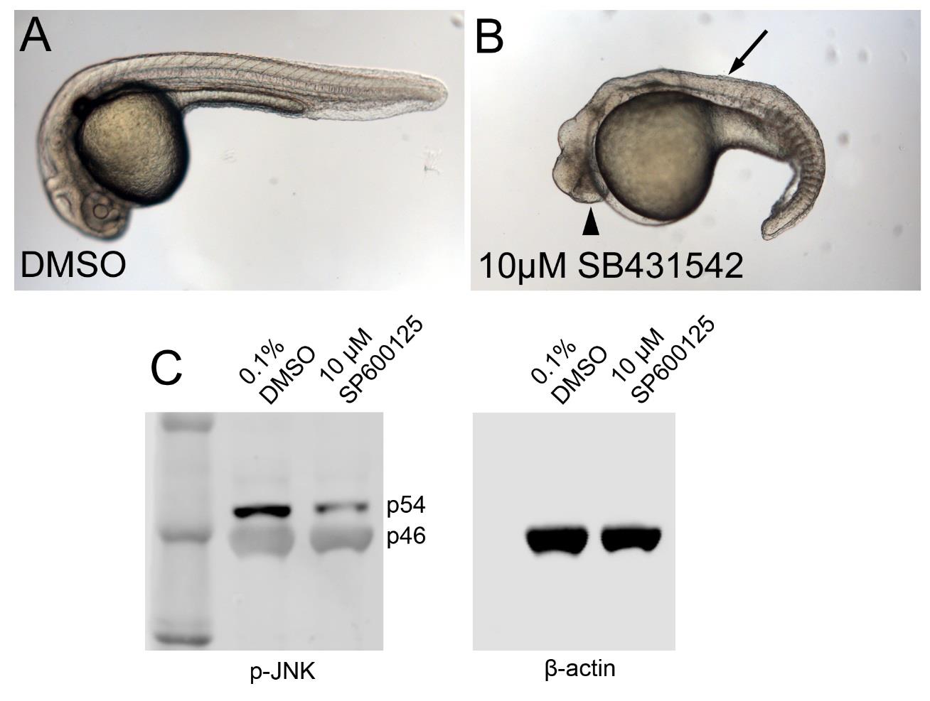
Fig. S5
Efficacy of SB-431542 (TGFβ) and SP600125 (JNK) inhibitors. (A,B) Live images of embryos treated with 0.1% DMSO (A, control) or with 10 µM SB431542 (B), starting at 4 hours post fertilization (hpf); 24 hpf, lateral view. The SB431542-treated embryo displays fused eyes (arrowhead) and lack of trunk somites (arrow), resembling the phenotype of mutants lacking the TGFβ family members Ndr1 and Ndr2 (Feldman et al., 1999), indicating that 10 µM SB431542 efficiently blocks TGFβ signalling (n=45/48; 2 independent experiments). (C) Duplex anti-pJNK (1:1000, Cell Signaling Technology, 4668s) and anti-&beta-actin (loading control; 1:1000, Sigma-Aldrich, A2228) Western blot of protein extracts from fins of adult zebrafish, after treatment with 0.1% DMSO (control) or 10 µM SP600125 for 5 hours, and separation via 9% SDS-PAGE. Levels of the phosphorylated version of the p54 splice isoform of JNK are several-fold reduced in the SP600125-treated sample. Similar results were obtained in three independent experiments.