
|
Fig. 6
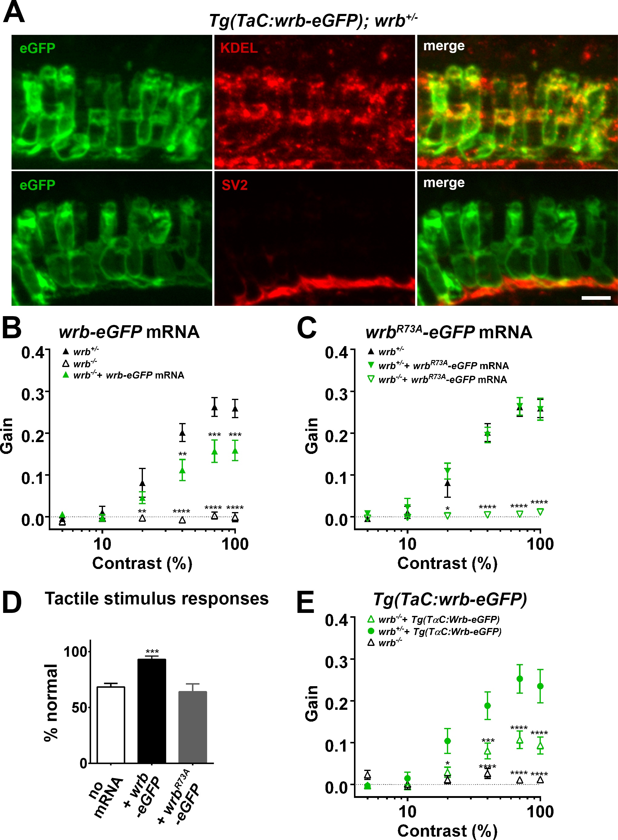
Photoreceptor wrb expression is critical for normal visual sensitivity. (A) Immunofluorescent images 10 µm-thick transverse cryosections of 5 dpf retinas from Tg(TaC:wrb-eGFP) stained antibodies to GFP (green), KDEL (top, red) or SV2 (bottom, red) to label ER and synapse respectively. (B) OKR gain versus log contrast plots for 5 dpf wrb+/- (closed triangles), wrb-/- mutants (open triangles), or wrb-/- mutants injected with mRNA encoding wrb-eGFP (green triangles). (C) Gain of OKR versus log contrast plots for 5 dpf wrb+/- heterozygous larvae (closed triangles), wrb+/- larvae injected with mRNA encoding wrbR73A-eGFP (green closed triangles), or wrb-/- mutants injected with mRNA encoding wrbR73A-eGFP (green open triangles). (D) Quantification of the percentage of larval offspring from a wrb heterozygous mating showing normal avoidance responses to light tail touch. Roughly 25% of larvae offspring (homozygous wrb-/- mutants) failed to show normal responses. Almost 100% of larvae exhibited normal touch responses following injection of mRNA encoding wrb-eGFP into 1-cell embryos. 25% of larvae failed to show a normal response following injection of mRNA encoding wrbR73A-eGFP. (E) OKR gain versus log contrast plots from 5 dpf heterozygous and homozygous wrb mutants carrying the Tg(TαC:wrb-eGFP) transgene. *P < 0.05. **P < 0.01. ***P < 0.001. ****P < 0.0001. Scale bar: in (A): 5 µm.