
|
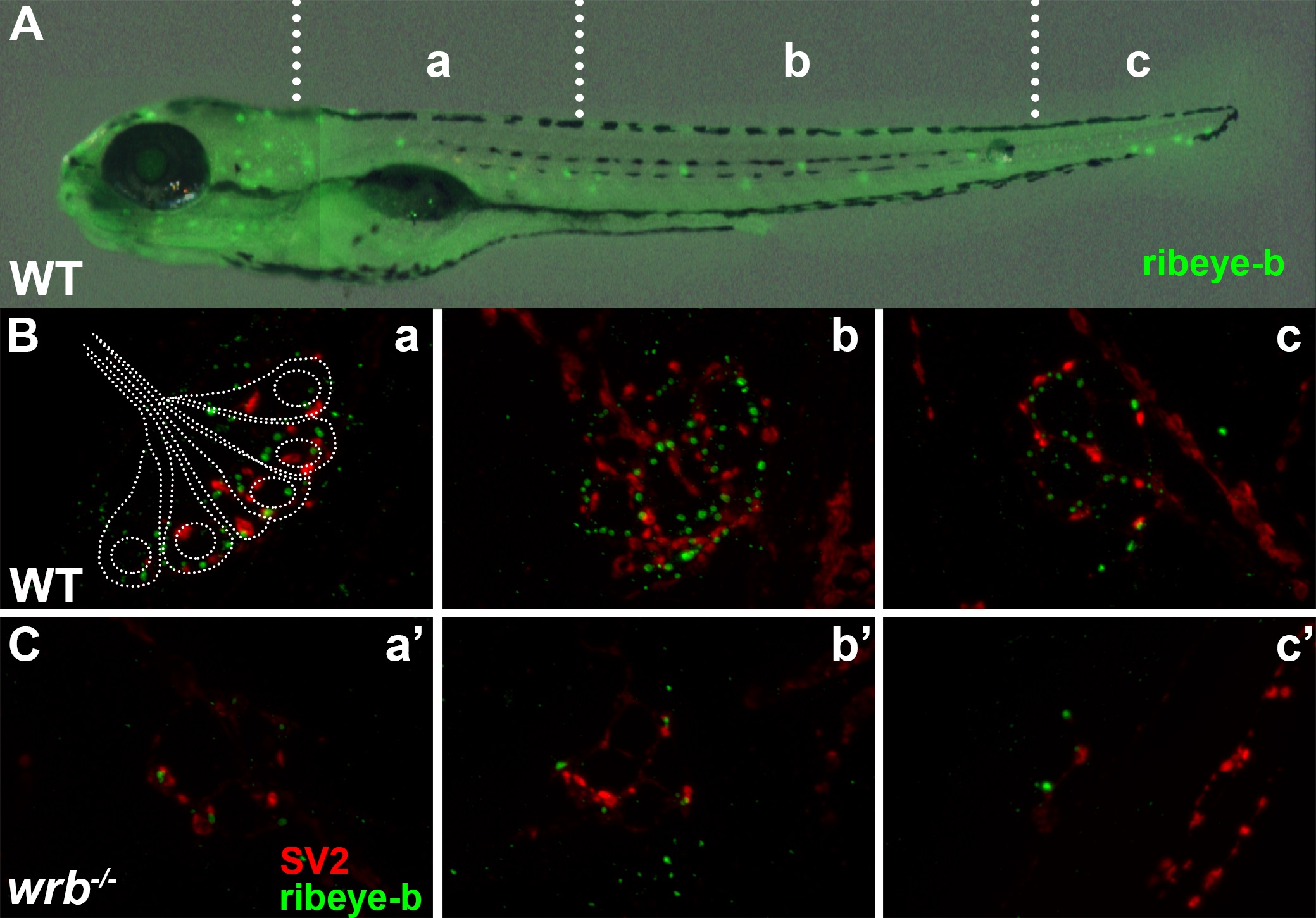
Fig. 4
Loss of Wrb disrupts endogenous ribeye localization at synaptic ribbons in hair cells at 5 dpf. (A) Lateral image of 5 dpf larvae immunostained with ribeye b antibodies to label lateral line hair cells. Hair cells in rostral (a, a′), middle (b, b′), and caudal (c, c′) regions of the trunk were imaged by fluorescence microscopy. (B, C) Fluorescence images showing individual neuromasts immunolabeled with SV2 (red) and ribeye-b (green) antibodies in wild-type and wrb-/- mutant larvae. Dotted outline in (B) depicts the orientation of individual hair cells within a single neuromast, as it relates to synaptic immunoreactivities.