
|
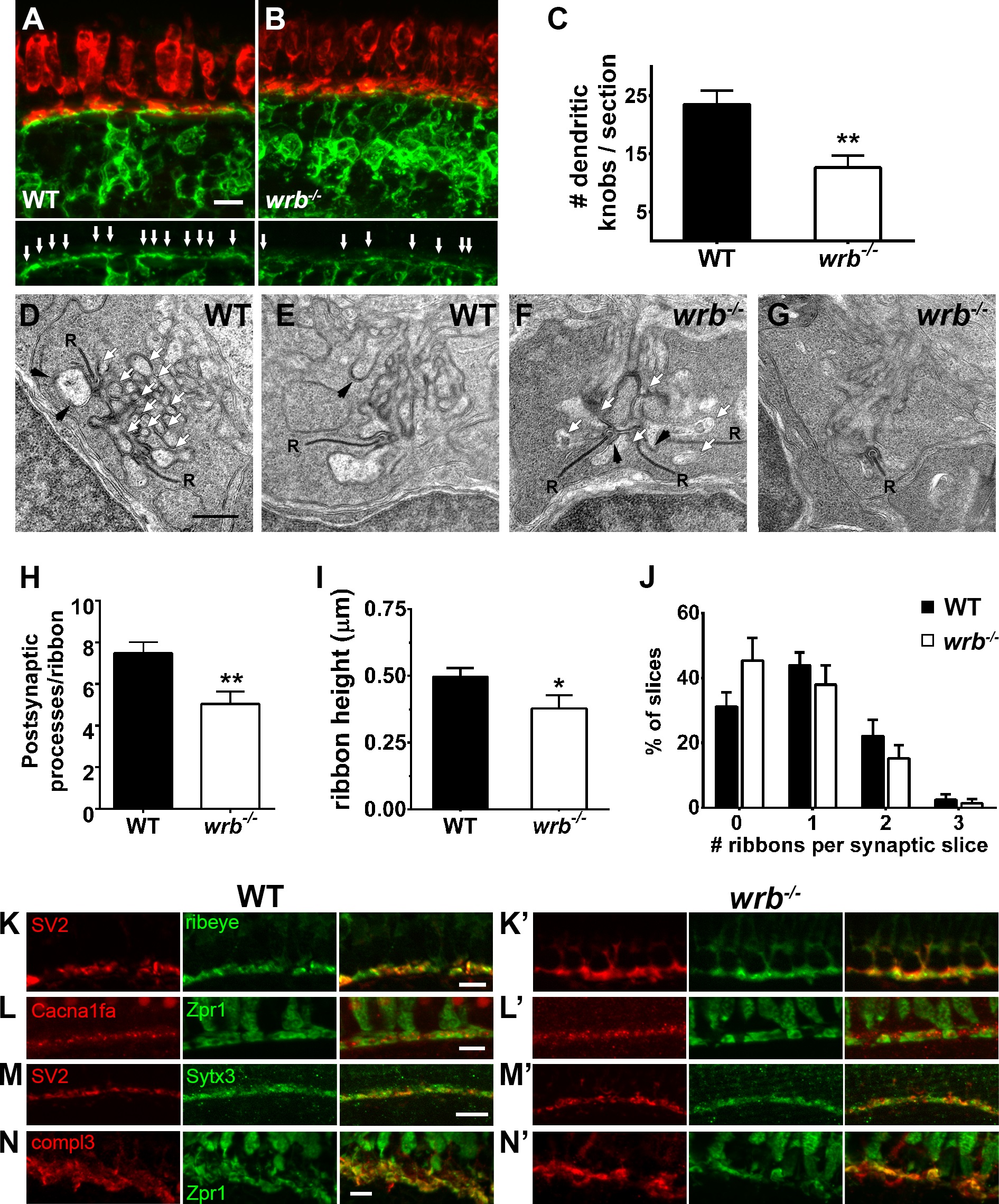
Fig. 3
Presynaptic and postsynaptic alterations in wrb-/- mutant photoreceptors. (A, B) Representative images of the OPL from cryosections of 5 dpf Tg(nyx:mYFP) and wrb-/-; Tg(nyx:mYFP) mutant retinas immunostained for red/green double cones (zpr1, red) and GFP (green). Bottom panels show bipolar dendritic projections (white arrows) within the OPL at higher magnification. (C) Quantification of dendritic invaginations inside cone pedicles across several cryosections (n = 4 and 8 sections for wild type and wrb-/-, respectively). (D-F) Transmission electron microscopy images of cone pedicles. Synaptic ribbons (R) were surrounded by postsynaptic processes (white arrows denote representative processes). Horizontal cells could be identified by characteristic densities and electron-lucent cytoplasm (black arrowheads). Quantification of postsynaptic processes per ribbon in wild-type and mutant cone pedicles (n = 26 wild-type, n = 19 wrb-/- synapses). (I) Quantification of photoreceptor ribbon heights in wild-type and mutant synapses (n = 32 wild-type, n = 18 wrb-/- synapses). (G) Quantification of average number of synaptic terminals in which 0, 1, 2, or 3 ribbons were encountered (n = 163 wild-type, n = 216 wrb-/- synapses). (H-K′) Immunohistochemistry of 5 dpf retinal cryosections with indicated photoreceptor presynaptic markers. Images were centered at the OPL of wild-type and wrb-/- mutants. Cacna1fa, pore forming alpha subunit of the presynaptic L-type calcium channel; SV2, synaptic vesicle protein 2; Sytx3, photoreceptor-specific target SNARE syntaxin 3B, compl3- exocytosis regulator complexin 3. * P < 0.05. **P < 0.001. Scale bars: 5 µm (A, B, H-I′, K, K′); 10 µm (J, J′); and 0.5 µm (D-G).