
|
Fig. 4
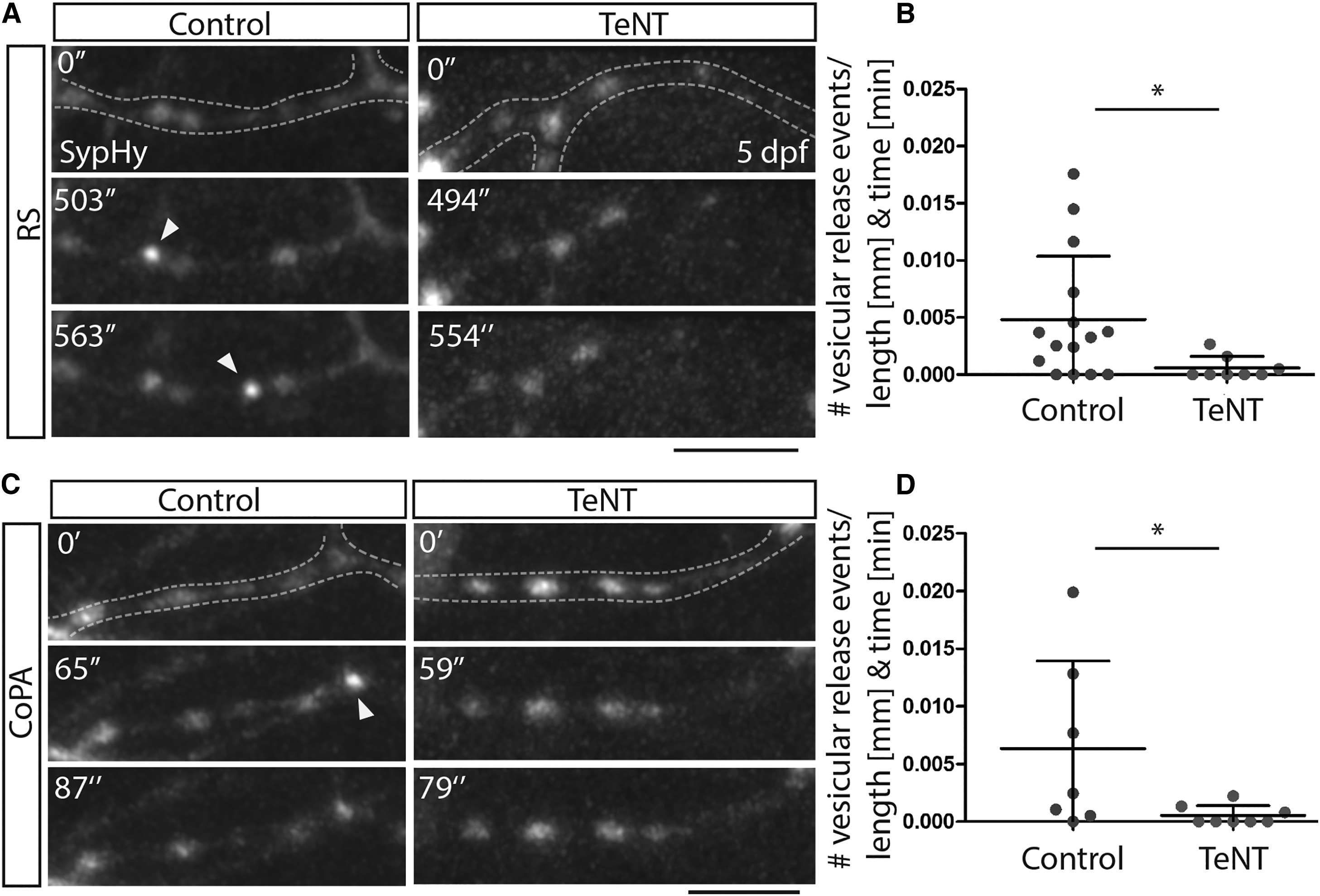
Tetanus Toxin Expression in Reticulospinal and CoPA Neurons Impairs Vesicular Release from Their Axons
(A) Images from time-lapse movies of sypHy expression in reticulospinal axon collaterals in control (left) and TeNT-expressing (right) neurons at 5 dpf. Dashed lines outline the collateral. Arrowheads point to punctate increases in GFP expression indicative of vesicular release. Scale bar, 5 µm.
(B) Quantitation indicates number of GFP events per collateral per micron per minute in control and TeNT-expressing reticulospinal neurons.
(C) Images from time-lapse movies of sypHy expression in CoPA axon collaterals in control (left) and TeNT-expressing (right) neurons at 5 dpf. Dashed lines outline the collateral. Arrowheads point to punctate increases in GFP expression indicative of vesicular release. Scale bar, 5 µm.
(D) Quantitation indicates number of GFP events per collateral per micron per minute in control and TeNT-expressing CoPA neurons.
See also Movies S1, S2, S3, and S4.