
|
Fig. 2
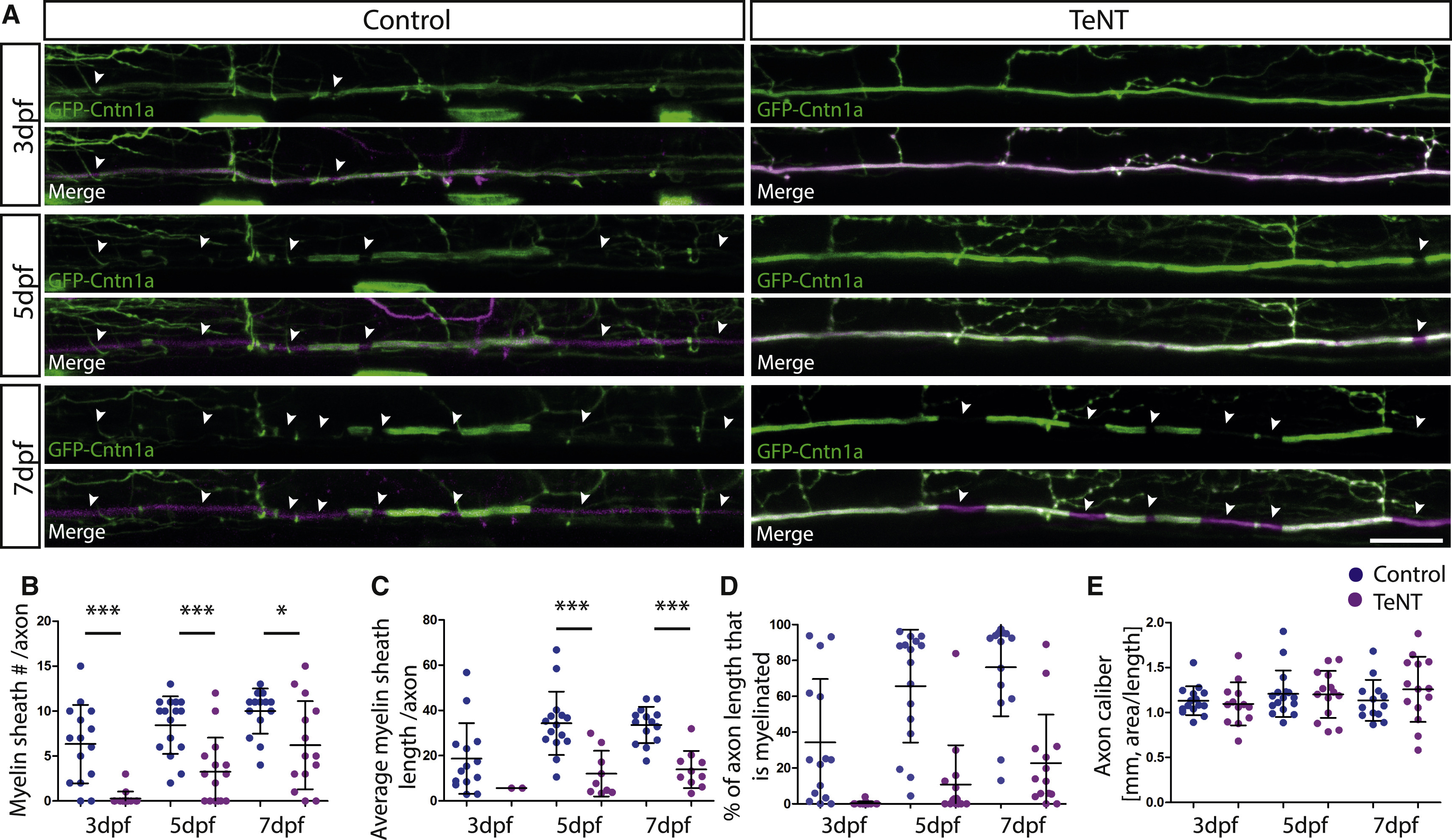
TeNT Expression in Reticulospinal Neurons Impairs Myelination along Individual Axons
(A) Individual reticulospinal axons labeled with GFP-Cntn1a and TdTomato (left) and with GFP-Cntn1a and TeNT-Tdtomato (right) at 3 dpf, 5 dpf, and 7 dpf. Scale bar, 15 µm.
(B-E) Quantification of myelin sheath number per axon per 425-µm imaging window (B), average length of myelin sheath per axon (C), percentage of axon length (per 425-µm imaging window) that is myelinated (D), and axon caliber (E) at 3 dpf, 5 dpf, and 7 dpf in control and TeNT-expressing reticulospinal neurons. All error bars indicate ± SD. See also Figure S3.