
|
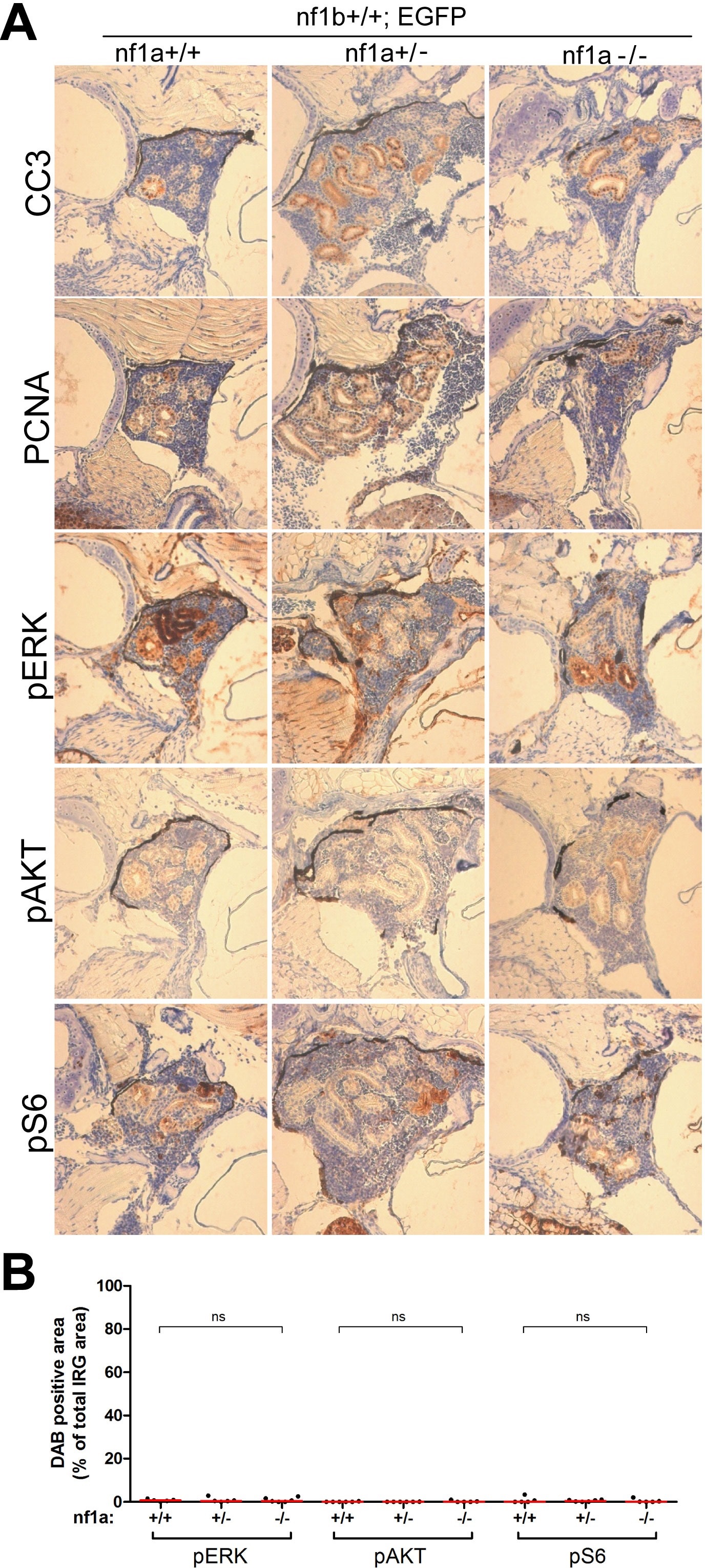
Fig. S6
No aberrant ERK/AKT/mTOR signaling was detected in the IRG of nf1a mutant fish that lacked MYCN overexpression.
(A) Immunohistochemical analysis of sagittal sections through the IRG of nf1a+/+;nf1b+/+;GFP, nf1a+/-;nf1b+/+;GFP and nf1a-/-;nf1b+/+;GFP fish at the age of 6 weeks, using antibodies against phosphorylated ERK1/2 (pERK), phosphorylated AKT (pAKT), phosphorylated S6 (pS6), cleaved Caspase-3 (CC3) and proliferating cell nuclear antigen (PCNA). The quantification of pERK-, pAKT- and pS6-positive IRG areas are shown in (B), with the red bars representing the median values. ns p>0.05, *p<0.05, **p<0.01 by two-tailed unpaired t-test.