
|
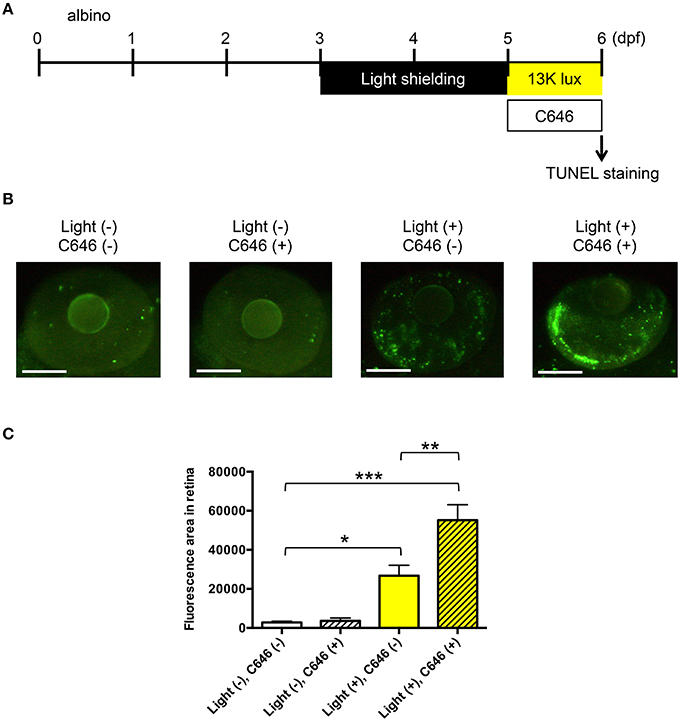
Fig. 4
Inhibition of EP300 increases retinal apoptosis in a larval zebrafish model of light-induced retinopathy. (A) Protocol for light-induced retinal damage in larval zebrafish. Zebrafish are shielded from light between 3 and 5 days post-fertilization (dpf) and then exposed to normal conditions (14 h 250 lux/10 h dark) or intense light (13,000 lux) in the presence or absence of 2 µM C646 for 24 h at 27°C. After light exposure, whole-mount TUNEL staining was performed. (B) Representative images of TUNEL staining in the retina of zebrafish exposed to intense light (indicated as light+) or normal light conditions (light-). Scale bars, 100 µm. (C) Quantitative analysis of retinal apoptosis in zebrafish exposed to the conditions shown in (B). *p < 0.01, **p < 0.001, ***p < 0.0001. Data are the mean ± SEM of 13-14 zebrafish/group.