
Image
|
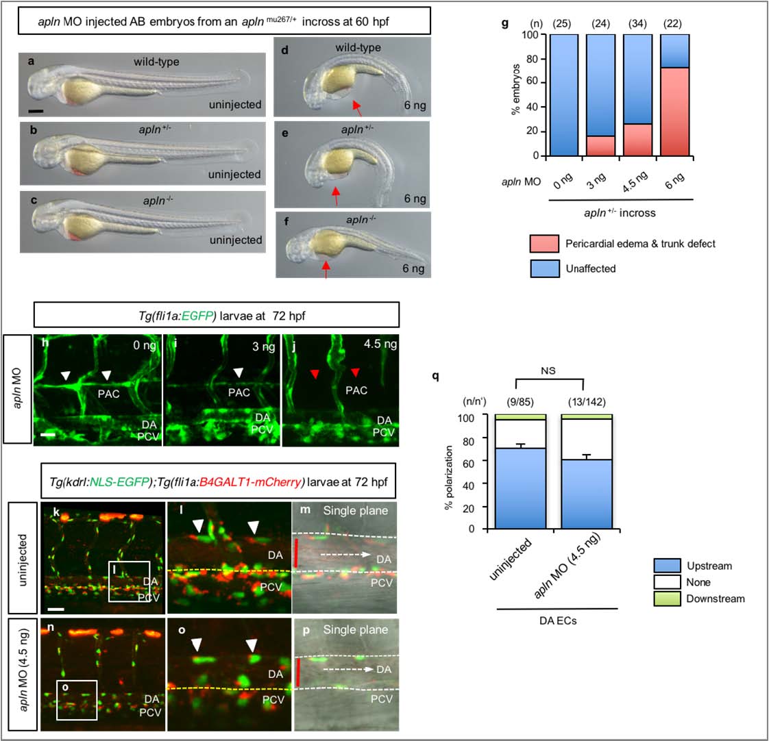
Figure Caption
Fig. S6
Endothelial cell polarization appears unaffected in apln morphants.
(a-f) Bright field images of 60 hpf embryos from an apln+/- incross uninjected (a-c) or injected with apln MO (6 ng) (d, e, f). Red arrows point to pericardial edema. (g) Quantative analysis of 60 hpf apln MO injected embryos from an apln+/- incross. The numbers of larvae (n) are indicated above the graph. (h-j) 3D rendered confocal images of 72 hpf Tg(fli1a:EGFP) larvae injected with apln MO (4.5 ng). White arrowheads point to the PAC vessel (h, i). Red arrowheads indicate absence of PAC in apln morphants (j). (k-p) Confocal images (lateral views) of 72 hpf Tg(kdrl:NLS78 EGFP);Tg(fli1a:B4GALT1-mCherry) larvae uninjected (k-m) or injected (n-p) with apln MO(4.5 ng). The white boxes in the left panels (k, n) are enlarged in the middle panels (l, o) and the single plane images, with brightfield, of those boxed areas are enlarged in (m) and (p). (q) Quantitative analysis of EC polarization in 72 hpf uninjected and apln MO injected (4.5 ng) larvae. The numbers of larvae (n) and ECs (n′) are indicated above the graph. Anterior to the left and dorsal to the top. Scale bars, 100 µm (a-f), 20 µm (h-p). DA, dorsal aorta; PCV, posterior cardinal vein. Error bars, SEM.
Figure Data
Acknowledgments
This image is the copyrighted work of the attributed author or publisher, and
ZFIN has permission only to display this image to its users.
Additional permissions should be obtained from the applicable author or publisher of the image.
Full text @ Nat. Commun.